Category:Stomach and Abomasum - Inflammatory Pathology
Stomach and Abomasum - Inflammatory Pathology
Gastritis refers to inflammation of the stomach.
Content
Gastritis, Catarrhal
Oedema Disease In The Pig
Fibrinous/ Diptheric Gastritis
- Not very common, but has severe consequences.
- Dirty-white, crumbly fibrin is seen on the surface of mucosa.
- Causes
- Toxic
- From drinking battery acid or other caustic material.
- Also gives with stomatitis and oesophagitis as well.
- Poisons such as mercuric chloride and carbolic acid also cause fibrinous/ diptheric gastritis.
- From drinking battery acid or other caustic material.
- Severe systemic disease
- e.g. septicaemic Erysipelas and Swine Fever in pigs, or septicaemic Salmonellosis.
- Not usually a primary problem but part of more severe generalised disease problem.
- Toxic
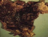
Haemorrhagic Gastritis
Clinical
- Usually only seen post mortem.
- Stomach full of thick tarry clots.
- Occasionally will vomit blood in life.
Pathology
Gross
- Wall of stomach is blacked and ulcerated.
- Red, thickened, necrotic, haemorrhagic mucosa.
Histologically
- Coagulative necrosis with fibrin, oedema, haemorrhage, and sometimes emphysema.
- May extend deep into submucosa/muscle.
Pathogenesis
- There are several causes of haemorrhagic gastritis
- Aspirin and non-steroidal anti inflammatory drug toxicity.
- Peracute / acute infections, e.g.
- Swine Fever
- Anthrax
- Leptospirosis in dogs (Leptospira icterohaemorrhagiae).
- Clostridial disease
- e.g. Braxy (Clostridium septicum)
- Affects older lambs or yearlings producing sudden death.
- Usually seen on sheep grazing on frosted grass so more common in colder areas.
- Bacterial exotoxin causes acute abomasitis.
- Pathology- At post mortem the stomach is grossly distended with partially clotted blood. The wall of the stomach is thickened,reddened and oedematous.
- Diagnosed by isolation of organism from the stomach wall.
- Is now usually vaccinated against (Heptovac 7 in 1 clostridial vaccine).
- e.g. Braxy (Clostridium septicum)
- Warfarin poisoning.
Vesicular Gastritis
- Is not seen, as the stomach has no stratum spinosum.
Chronic gastritis
- Chronic gastritis is usually proliferative rather any other type of gastric inflammation.
- Usually a parasitic cause.
- Occurs mostly in the pig and in cattle.
- Pig
- Redworms (Hyostrongylus)
- Seen mostly in sows, and are present in up to 30% of pig herds.
- Small numbers produce little pathology, but large numbers cause thin sow syndrome.
- Animals eat well but slowly lose condition.
- Cattle
- Ostertagiasis produces a condition similar to thin sow syndrome.
Chronic Hypertrophic Gastritis In The Dog
- Clinically see anorexia, weight loss, anaemia and associated hepatic disease.
- Associated with protein loss into gut.
Pathology
- Hyperplasia of mucosa.
- Mucosa thrown up into folds.
- Reduced numbers of parietal cells and increased numbers of goblet cells.
Chronic Atrophic Gastritis In The Dog
- Aetiology uncertain.
- Grossly: (may be difficult to appreciate)
- Reduced mucosal thickness.
- Loss of rugae.
- Histologically
- Mucosal thinning.
- Loss of gastric glands.
- Diffuse inflammatory infiltrate of lymphocytes and plasma cells.
- Fewer eosinophils in lamina propria.
Pages in category "Stomach and Abomasum - Inflammatory Pathology"
The following 10 pages are in this category, out of 10 total.
