Cryptosporidium
Jump to navigation
Jump to search
| This article has been peer reviewed but is awaiting expert review. If you would like to help with this, please see more information about expert reviewing. |
Recognition
- Minute protozoan parasite
- Wide host range
- Parasitises epithelial cells lining the alimentary and respiratory tracts
- Developmental stages confined to the microvillous brush border
- C. parvum most associated with disease in domestic animals and in humans
- Other species affect birds
- Small oocysts of 4-5μm
Life Cycle
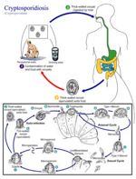
- Direct life cycle
- Only one host
- Homoxenous
- 1 week prepatent period
- Sporulated oocysts passed in faeces
- Autoinfection can occur
- Thin walled oocysts
- Faecal-oral transmission also occurs
- Thick walled oocysts
Pathogenesis
- Causes outbreaks of diarrhoea in young animals
- Common cause of calf-hood scours
- Older animals may be asymptomatic carriers
- Contributes to undifferentiated neonatal calf diarrhoea which is a mixed viral enteritis in calves
- Common infection in AIDS patients
Epidemiology
- Direct faecal-oral infection
- E.g. School parties visiting farms
- Water-borne infection
- E.g. contaminated water supply may infect hundreds of people
- Difficult to locate source
Diagnosis
- Faecal smear
- Ziehl-Neelson (ZN) stain
- Oocysts stain red against a blue/green background
- Immunoassays
- Detect oocysts in faeces
Control
- Isolate/quarantine bought-in calves
- Treat if signs of diarrhoea present
- Good hygiene, adequate bedding and disinfection of calf pens is important
- Prevention/treatment
- Halofuginone
- Halocur or Intervet
- Oral dosage
- Halofuginone
Villus Atrophy in Enteritis
- Affects calf, lamb, piglet, kitten.
- Increasingly important as part of the neonatal diarrhoea complex in calves.
- Zoonosis.
Pathology
Gross
- Intestines diffusely reddened, with fluid contents.
Histological
- Tiny parasites on surface of epithelium.
- Villus atrophy and fusion.
- Iinflammation (mainly lymphoid) in crypts and lamina propria.


