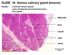
Serous Salivary Gland - Anatomy & Physiology
Jump to navigation
Jump to search
Overview
The serous salivary gland has a connective tissue capsule and septa dividing the parenchyma into lobes. There is a duct system. Interlobular ducts run in the tissue septum lined by cuboidal to columnar epithelium.
Intralobular ducts run within the lobules. Striated intralobular ducts lined with cuboidal epithelium. Intercalated intralobular ducts lined with low cuboidal to simple squamous epithelium. Serous acini secrete a watery solution rich in proteins with spherical nuclei. Cells are pyramidal, cuboidal or crescent shaped.
