Difference between revisions of "Bursa of Fabricius"
m (Text replace - "[[Lymphocytes|" to "[[Lymphocytes - Introduction|") |
Fiorecastro (talk | contribs) |
||
| (4 intermediate revisions by 2 users not shown) | |||
| Line 1: | Line 1: | ||
| + | |||
[[Image:LH_Bursa_Photo.jpg|thumb|150px|right|<p>'''Bursa of Fabricius'''</p><sup>©Nottingham Uni 2008</sup>]] | [[Image:LH_Bursa_Photo.jpg|thumb|150px|right|<p>'''Bursa of Fabricius'''</p><sup>©Nottingham Uni 2008</sup>]] | ||
==Introduction== | ==Introduction== | ||
| Line 7: | Line 8: | ||
<p>Lymphoid precursors migrate into the developing bursa during the first few weeks of embryo development. The bursa continues to grow through development and in chickens the bursa reaches its maximum size by six weeks. After this it slowly regresses (involutes) until only a small remnant is present in the adult.</p> | <p>Lymphoid precursors migrate into the developing bursa during the first few weeks of embryo development. The bursa continues to grow through development and in chickens the bursa reaches its maximum size by six weeks. After this it slowly regresses (involutes) until only a small remnant is present in the adult.</p> | ||
==Structure== | ==Structure== | ||
| − | <p>The bursa is a round out-pouching of the [[ | + | <p>The bursa is a round out-pouching of the [[Avian Vent and Cloaca - Anatomy & Physiology|cloaca]]. It is found on the caudodorsal surface of the [[Avian Vent and Cloaca - Anatomy & Physiology|cloaca]] (in the proctodeum) cranial to the dorsal proctodeal gland. The bursa consists of a number of lymphoid lobules and crypt-like folds surrounding a lumen, which is enclosed in a thin layer of stratified squamous epithelium. The lumen opens into the proctodeum.</p> |
<p>Like the [[Thymus - Anatomy & Physiology|thymus]] the lobules have a cortex and medulla and the [[Lymphocytes - Introduction|lymphocytes]] are supported by epithelial cells. B lymphocytes developing from the lymphoid precursors gather in the cortex, hence the cortex stains stronger than the medulla.</p> | <p>Like the [[Thymus - Anatomy & Physiology|thymus]] the lobules have a cortex and medulla and the [[Lymphocytes - Introduction|lymphocytes]] are supported by epithelial cells. B lymphocytes developing from the lymphoid precursors gather in the cortex, hence the cortex stains stronger than the medulla.</p> | ||
===Histology=== | ===Histology=== | ||
| Line 17: | Line 18: | ||
<p>The bursa’s primary function is maturating and causing the differentiation of [[Lymphocytes#B cells|B lymphocytes]], it also produces the hormone bursin which activates [[Lymphocytes#B cells|B lymphocytes]]. </p> | <p>The bursa’s primary function is maturating and causing the differentiation of [[Lymphocytes#B cells|B lymphocytes]], it also produces the hormone bursin which activates [[Lymphocytes#B cells|B lymphocytes]]. </p> | ||
<p>The bursa has some [[Lymphocytes#T cells|T lymphocytes]] present near to its opening into the proctodeum where they survey antigens.</p> | <p>The bursa has some [[Lymphocytes#T cells|T lymphocytes]] present near to its opening into the proctodeum where they survey antigens.</p> | ||
| + | <br> | ||
| + | {{Template:Learning | ||
| + | |OVAM=[http://www.onlineveterinaryanatomy.net/content/histology-avian-bursa-fabricius Bursa of Fabricius Histology] | ||
| + | }} | ||
| + | <br><br> | ||
| + | {{Jim Bee 2007}} | ||
| − | + | ==Webinars== | |
| + | <rss max="10" highlight="none">https://www.thewebinarvet.com/internal-medicine/webinars/feed</rss> | ||
[[Category:Primary Lymphoid Tissue]] | [[Category:Primary Lymphoid Tissue]] | ||
Latest revision as of 15:16, 5 January 2023

Introduction
Also called the cloacal bursa
It is a primary lymphoid organ found in birds. The bursa was the first place that a certain subset of lymphocytes was observed and consequently they were named B lymphocytes (bursa of Fabricius or bursa equivalent organs). The bursa is involved in the differentiation of B lymphocytes.
Development

Lymphoid precursors migrate into the developing bursa during the first few weeks of embryo development. The bursa continues to grow through development and in chickens the bursa reaches its maximum size by six weeks. After this it slowly regresses (involutes) until only a small remnant is present in the adult.
Structure
The bursa is a round out-pouching of the cloaca. It is found on the caudodorsal surface of the cloaca (in the proctodeum) cranial to the dorsal proctodeal gland. The bursa consists of a number of lymphoid lobules and crypt-like folds surrounding a lumen, which is enclosed in a thin layer of stratified squamous epithelium. The lumen opens into the proctodeum.
Like the thymus the lobules have a cortex and medulla and the lymphocytes are supported by epithelial cells. B lymphocytes developing from the lymphoid precursors gather in the cortex, hence the cortex stains stronger than the medulla.
Histology
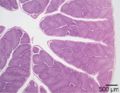
Low power magnification
©Nottingham Uni 2008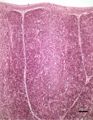
High power magnification
©Nottingham Uni 2008


Functions
The bursa’s primary function is maturating and causing the differentiation of B lymphocytes, it also produces the hormone bursin which activates B lymphocytes.
The bursa has some T lymphocytes present near to its opening into the proctodeum where they survey antigens.
| Bursa of Fabricius Learning Resources | |
|---|---|
Anatomy Museum Resources |
Bursa of Fabricius Histology |
| Originally funded by the RVC Jim Bee Award 2007 |
Webinars
Failed to load RSS feed from https://www.thewebinarvet.com/internal-medicine/webinars/feed: Error parsing XML for RSS