CNS Development - Anatomy & Physiology
Introduction
Development of the Central Nervous System (CNS) includes development of the brain, spinal cord, optic and auditory systems, as well as surrounding supporting cells including ependymal cells, astrocytes, oligodendrocytes and microglia. Information within this page will exclude development of the Peripheral Nervous System (PNS) which includes nerve and ganglia formation. For further information on PNS development, please see the PNS development page.
For the appropriate development of the CNS a number of important basic requirements must be achieved. For example, the appropriate haemopoeitic cells must differentiate into specific cells (either neurons or glia) and any axons must extend to appropriate positions in order to form synapses. Development of the CNS is a complex process and complete coordination is required to facilitate the correct procedure of cell differentiation and the establishment of the correct intracellular connections. A range of neurotrophic factors regulate these interactions including FGF, nerve growth factor, laminin, brain derived neurotrophic factor and neurotrophin 3.
Developmental Origins
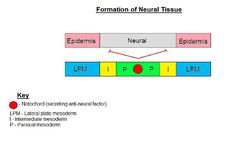
The embryological origin of the CNS is from the neural ectoderm which is a ridge of tissue in the centre of the early embryo once the basic blastula has undergone a degree of differentiation. The neural ectoderm is formed via the thickening of the ectoderm and it's interaction with underlying basic neural tissues including the notochord. This interaction results in the formation of the neural plate, termed the neuroectoderm. Below is a diagram of the completed initial stages of development to provide an indication of the anatomy of the development process.
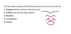
Formation of the neuroectoderm occurs as a result of the folding of the neural plate due to the presence of the Lateral Plate Mesoderm (LPM) factor. The edges of the neural plate begin to fold upwards and inwards, eventually leading to the formation of a neural crest and tube. LPM also initiates the formation of epidermis from ectoderm and therefore areas of ectoderm dorsal to the neural tube differentiate into epidermis.
Neural Differentiation
In the early embryo as shown in the images above, the neural tube is formed and represents the initial stages of the formation of the brain and spinal cord. As the embryo develops, the neural tube continues to develop in conjunction with the formation of the vertebrae of the spine.
Development of Vertebrae
Formation of the cells that will be the precursors to the spinal vertebrae align in an intersegmental fashion to the neural cells of the spinal cord, in a form of bridging pattern. Therefore each neural cell has a spinal vertebrae precursor cell between itself and the adjacent neural cell such that the spinal cell effectively overlaps the edges of two neural cells. This ensures that as the spine and spinal cord develop the spinal nerves become aligned to the spaces between the vertebrae that will eventually become seperated by intervertebral discs.
Further along into spinal development, each vertebral body develops three centres of ossification which are separated by cartilage growth plates. Each half of the two vertebral arches that surround the developing spinal cord (neural tube) have their own ossification centre facilitating the development of vertebral processes, ribs and other bone structures related to the LPM including the scapula and pelvis.
Development of the Brain
Development of the brain begins with a series of expansions of the neural tube at the cranial end, giving rise to the internal spaces of the brain, the ventricles and aqueducts. The initial expansion of the neural tube is in the mesencephalon area, or mid brain area. This is then followed by expansions in the rhombencephalon (hind brain) and in the proencephalon (forebrain).
After these initial expansions, a series of further developments occurs to form some of the major landmarks in the brain. The mesencephalon narrows prior to the proencephalon resulting in the formation of the 'Foramen of Munro' and the 'Cerebral Aqueduct'. The proencephalon develops two further pouches in the cranial lateral aspect which are the foundations of the 'Lateral Ventricles' and the 'Third Ventricle'.
Caudal Spinal Cord Development
In late development, the spinal cord extends along the entire length of the spinal canal (formed by the spinal vertebrae). The spinal cord then terminates at the 'conus medullaris' in the middle of the lumbar spine. In adults the spinal cord ends at the beginning of the lumbar psine. Lumbar and sacral nerves extend via the 'cauda equine'.
Therefore in neonates CSF taps must be caudal to the conus medullaris to avoid damaging the spinal cord.
Origins of Functional Types of Neurone
| Nerve | Origin |
|---|---|
| Sensory - Afferent | Neural crest |
| Motor - Efferent | Basal plate of neural tube |
| Association - Interneurones | Alar plate of neural tube |
| This article is still under construction. |


