Difference between revisions of "Duodenum - Anatomy & Physiology"
Jump to navigation
Jump to search
| Line 16: | Line 16: | ||
==Vasculature== | ==Vasculature== | ||
| − | * The duodenum recieves blood from: | + | *The duodenum recieves blood from: |
**Coeliac artery | **Coeliac artery | ||
| − | **Cranial | + | **Cranial mesenteric artery |
*Both are branches of the aorta. | *Both are branches of the aorta. | ||
| + | *The cranial mesenteric vein drains blood from the duodenum into the portal vein. | ||
| + | **This blood, carrying the products of digestion, enters the liver. | ||
==Histology== | ==Histology== | ||
Revision as of 14:48, 10 July 2008
Introduction
The duodenum is the proximal part of the small intestine. It extends from the pylorus of the stomach to the jejunum. Both the pancreatic and bile ducts open into the duodenum.
Structure
- It has descending and ascending portions.
- The descending duodenum passes out of the pylorus of the stomach (on the right side of the abdomen) and has a sigmoid flexure. It passes towards the right abdominal wall and rises dorsally. In its passage it is related dorsally to the right lobe of the pancreas, ventrally to the jejunum and medially to the ascending colon and caecum.
- At a point between the right kidney and pelvic inlet it turns medially and cranially around the root of the mesentry to become the ascending duodenum. The point of turn is called the caudal flexure of the duodenum.
- The ascending duodenum is shorter and bends ventrally to enter the mesentery and becomes the jejunum.
- Mesoduodenum attaches the duodenum to the dorsal abdominal wall. This is relatively short in the horse and ruminant and longer in the carnivore and pig.
Vasculature
- The duodenum recieves blood from:
- Coeliac artery
- Cranial mesenteric artery
- Both are branches of the aorta.
- The cranial mesenteric vein drains blood from the duodenum into the portal vein.
- This blood, carrying the products of digestion, enters the liver.
Histology
- The mucosa is arranged into villi that provide a large surface area for absorption.
- Epithelium is simple columnar - ideal for absorption.
- Epithelial cells are known as enterocytes.
- A single layer of enterocytes overlies the lamina propria.
- Enterocytes originate from progenitor cells that migrate from mucosal crypts. They differentiate as they migrate up the villus.
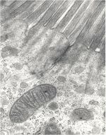
- Enterocytes ane absorptive and posses microvilli.
- Membrane bound enzymes and transport proteins are also within the epithelium.
- Each villus houses capillaries that transport amino acids, monosaccharides and other digestive products and lacteals that transport triacylglycerides. Lacteals drain into the lymphatic system.
- Crypts are present at the base of each villus in the mucosa. Cell types in mucosal crypts (from luminal to basal):
- goblet at the tip of the crypt. Produce mucous by exocytosis.
- entero-endocrine in the middle of the crypt. Secrete peptides and amines.
- paneth at the base of the crypt. Function unknown. Contain eosinophilic granules.
- Brunner's glands are present in the submucosa.
- Produce an alkaline secretion which neutralises stomach acid.
- Open into the crypts above in the mucosa.
- Lamina muscularis is smooth muscle.
- Contraction of smooth muscle shortens the villus. This helps to pump out absorbed products of digestion. Relaxation of smooth muscle lengthens the villus which increases surface area, facilitating absorption.

