|
|
| (40 intermediate revisions by the same user not shown) |
| Line 1: |
Line 1: |
| | {{unfinished}} | | {{unfinished}} |
| | ==Central Nervous System== | | ==Central Nervous System== |
| − | ===Brain=== | + | ===[[Equine Brain - Horse Anatomy|Brain]]=== |
| − | The brain is responsible for co-ordinating, integrating and controlling the rest of the nervous system. The brain is divided into several parts. Based on phylogenetic development, it can be divided into the forebrain, midbrain and hindbrain. Based on gross anatomy, it can be divided into the cerebrum, cerebellum and brainstem. The brain is enclosed within the cranial cavity of the skull.
| |
| − | ====Forebrain====
| |
| − | The ''forebrain (proencephalon)'' is the largest part of the brain, most of which is ''cerebrum''. Other important structures found in the forebrain include the ''thalamus'' , the ''hypothalamus'' and the ''limbic system''. The cerebrum is divided into two cerebral hemispheres connected by a mass of white matter known as the ''corpus callosum''. Each hemisphere is split into four lobes; the ''frontal'', ''parietal'', ''occipital'' and ''temporal'' lobes. The surface of each hemisphere is made up of grey matter known as the ''cerebral cortex'' and is folded to increase the surface area available within the skull. The cortex has roles within perception, memory and all higher thought processes. Inside the cortex is the ''white matter'', within which are a number of nuclei (grey matter), known as the ''basal nuclei''. The basal nuclei receive information from the cortex to regulate skeletal movement and other higher motor functions.
| |
| | | | |
| − | The thalamus functions to relay sensory information to the cerebral cortex and the hypothalamus, regulating visceral functions including temperature, reproductive functions, eating, sleeping and the display of emotion. The limbic system describes a collection of structures within the forebrain, including the ''amygdala'' and '' hippocampus'', also known as the 'emotional brain'. It is important in the formation of memories and in making decisions and learning.
| + | ===[[Equine Cranial Nerves - Horse Anatomy|Cranial Nerves]]=== |
| − | =====Thalamus===== | |
| − | The thalamus has many functions:
| |
| − | *Processing and relaying sensory information selectively to various parts of the cerebral cortex
| |
| − | *Translating signals to the cerebral cortex from lower centres including auditory, somatic, visceral, gustatory and visual systems
| |
| − | *Regulating states of sleep and wakefulness The thalamus plays a major role in regulating arousal, levels of consciousness and levels of activity.
| |
| | | | |
| − | =====Hypothalamus===== | + | ===[[Vasculature of the Equine Brain - Horse Anatomy|Vasculature of the Brain]]=== |
| − | The function of the hypothalamus is mainly related to the overall regulation of the [[:Category:Endocrine System - Anatomy & Physiology|Endocrine System]]. The hypothalamus is closely related to the [[Equine Endocrine System - Horse Anatomy#Pituitary Gland|pituitary gland]], controlling a large proportion of the activity going to it. For a more detailed analysis of the function of this part of the brain, please use the link: [[Hypothalamus - Anatomy & Physiology|Hypothalamus Anatomy and Physiology]].
| |
| | | | |
| − | =====Pituitary===== | + | ===[[Equine Spinal Cord - Horse Anatomy|Spinal Cord]]=== |
| − | The function of the pituitary is mainly related to the production of hormones as part of the Endocrine System. For further information on the pituitary gland please use this link: [[Equine Endocrine System - Horse Anatomy#Pituitary Gland|Equine Pituitary Gland]].
| |
| | | | |
| − | =====Cerebral Cortex===== | + | ===[[Equine Meninges - Horse Anatomy|Meninges]]=== |
| − | =====Limbic System=====
| |
| − | =====Olfactory Bulb=====
| |
| | | | |
| − | ====Midbrain==== | + | ===[[Equine Cerebrospinal Fluid - Horse Anatomy|Cerebrospinal Fluid]]=== |
| − | ====Hindbrain====
| |
| | | | |
| − | ===Cranial Nerves=== | + | ===Clinical Links=== |
| − | Cranial nerves arise from the brain and [[Hindbrain - Anatomy & Physiology|brain stem]], rather than the spinal cord. Nerves arising from the spinal cord are the [[PNS Structure - Anatomy & Physiology|peripheral nerves]]. There are 12 pairs of cranial nerves and these pairs of nerves passage through [[Skull and Facial Muscles - Anatomy & Physiology|foramina in the skull]], either individually or in groups. Cranial nerves are traditionally referred to by Roman numerals and these numerals begin cranially and run caudally.
| + | *[[Equine Protozoal Myeloencephalitis]] |
| − | The most cranial nerve is the '''Olfactory nerve (I)''' which runs from the nasal cavity through to the olfactory bulb. The next most cranial is the '''Optic nerve (II)''' which runs from the eyes to the [[Forebrain - Anatomy & Physiology#Thalamus|thalamus]]. Cranial nerves III to XII all exit from the brain stem and innervate the head, neck and organs in the thorax and abdomen. In order of most cranial to caudal, these include the '''Oculomotor nerve (III)''', the '''Trochlear nerve (IV)''', the '''Trigeminal nerve (V)''', the '''Abducens nerve (VI)''', the '''Facial nerve (VII)''', the '''Vestibulocochlear nerve (VIII)''', the '''Glossopharyngeal nerve (IX)''', the '''Vagus nerve (X)''', the '''Accessory nerve (XI)''' and the '''Hypoglossal nerve (XII)'''.
| + | *[[Equine Herpesvirus 1]] |
| | + | *[[Polyneuritis Equi]] |
| | | | |
| − | Many of the cranial nerves with nuclei within the brain stem contain sensory and motor neurone components. The sensory fibre components have their cell bodies located in ganglia outside the central nervous system and the motor fibre element have their cell bodies within the central nervous system. The'''Olfactory nerve (I)''', '''Optic nerve (II)''' and '''Vestibulocochlear nerve (VIII)''' are sensory nerves. The , '''Oculomotor nerve (III)''', '''Trochlear nerve (IV)''','''Abducens nerve (VI)''','''Accessory nerve (XI)''' and '''Hypoglossal nerve (XII)''' are motor nerves. Finally, the '''Trigeminal nerve (V)''', '''Facial nerve (VII)''','''Glossopharyngeal nerve (IX)''', and '''Vagus nerve (X)''' are mixed sensory and motor nerves.
| + | ==Peripheral Nervous System== |
| − | | + | ==Introduction== |
| − | ====Olfactory Nerve (I)==== | + | The '''Peripheral Nervous System''' is made up of cranial and spinal nerves. Spinal nerves are named after the vertebra immediately above it, except for '''cervical vertebra'''. There are '''7''' cervical vertebrae and '''8''' cervical spinal nerves. The peripheral nervous system can be divided into the '''somatic nervous system''' and '''autonomic nervous system'''. The somatic nervous system co-ordinates body movements and also receives external stimuli. It regulates activities that are under conscious control. The autonomic nervous system subdivided into the '''sympathetic nervous system''', '''parasympathetic nervous system''', and enteric division. The sympathetic nervous system is the '''‘fight or flight’''' system which comes into role when an animal is under threat, its main neurotransmitter is '''adrenaline'''. The parasympathetic nervous system is the '''‘rest and digest’''' system which is responsible for digestion. Its main neurotransmitter is '''acetylcholine'''. |
| − | The olfactory nerve is involved in the conscious perception of smell. Primary afferent cell bodies are located within the olfactory epithelium of the nasal mucosa on ethmoturbiate bones,rather than in a ganglion like the other cranial nerves. Projections from these cell bodies are the olfactory nerve fibres. The olfactory nerve is a sensory nerve and is composed of many '''Special Visceral Afferent''' fibres. The fibres are formed into bundles that are referred to as 'Olfactory filaments'. The olfactory nerve passes through the [[Skull and Facial Muscles - Anatomy & Physiology#Ethmoid Bone (os ethmoidale)|'''Cribiform plate''']] and is surrounded by meningeal sheets including the [[Meninges - Anatomy & Physiology#Subarachnoid_Space|sub-arachnoid space]]. The olfactory nerve terminates at the [[Forebrain - Anatomy & Physiology#Olfactory_Bulb|olfactory bulb]]. The horse also has nerves which arise from the nasal septum that course into the olfactory bulb, along with the '''vomeronasal nerve''' arising from the '''vomeronasal organ'''. Secondary neurons within the olfactory bulb project through the olfactory tracts to synapse with third order neurons in the medial forebrain bundle, amygdala, septal nuclei and habenular nuclei.
| + | ==Structure== |
| − | | + | [[Image:WIKIVETperipheralnervestructure.jpg|thumb|right|150px|Peripheral Nerve Structure. Sophie Stenner, RVC 2008]] |
| − | In the horse, special consideration must be given to diseases of the guttural pouch when considering cranial nerve dysfunction. The Glossopharangeal (IX), Vagus (X)and Accessory (XII) nerves are located in the medial compartment of the guttural pouch. The Facial (VII) nerve runs along the lateral compartment. The Mandibular nerve (V2) has limited contact with the dorsal wall of the lateral compartment.. Guttural pouch mycosis commonly results in paresis of cranial nerves IX,V and XII as well as erosion of the internal carotid artery. Rarely, there is involvement of cranial nerves VII and VIII.
| + | Nerve fibres reside in a connective tissue matrix called the '''endoneurium''' and are gathered together into bundles or fascicles defined by a second connective tissue layer called the '''perineurium'''. Groups of fascicles are then gathered together in a third connective tissue layer called the '''epineurium'''. Thus, peripheral nerves have a '''three-tiered hierarchical arrangement of connective tissue'''. '''Renaut bodies''' are loose, cigar-shaped whorls of extracellular matrix within fascicles that are common in equine nerves at points of stress or compression. |
| − | | + | ==Nerve Fibre== |
| − | ====Optic Nerve (II)====
| + | The nerve fibre consists of the impulse-carrying axon, which is surrounded by an ensheathing cell, the [[#The Schwann cell|Schwann cell]], which in turn is surrounded by an acellular basal lamina that is continuous along the length of the nerve. Nerve fibres come in various discrete diameter groups, which are reflected in their conduction velocities. The larger the diameter the more rapid the rate of impulse conduction. Particular targets or receptors are associated with axons of a particular diameter. Axons connected to muscles spindles have a large diameter (20 µm) and conduct at 120 m/s whilst the smallest myelinated fibres are about 1µm and conduct at around 6 m/s. The smallest fibres of all are the unmyelinated fibres (the high-threshold sensory afferents, or C-fibres, and post-ganglionic autonomies) and have a diameter of between 1 and 0.1 µm. These fibres do not conduct by saltatory conduction and have very slow conduction rates of around 0.5 m/s. |
| − | The optic nerve represents the connection between the receptor cells of the [[Eye - Anatomy & Physiology#The Wall (retina, uvea and sclera)|retina]] and the [[Forebrain - Anatomy & Physiology|forebrain]]. It is not a true nerve, but represents an extension of the brain. The optic nerve is sesory, and is composed of '''Special Somatic Afferent fibres'''.
| + | ===Axon=== |
| − | | + | Axons have an outer membrane called the '''axolemma''' and within this there is the '''axoplasm''' which is continuous with the cytoplasm of the [[Neurons - Anatomy & Physiology|neuron]]. There are no ribosomes, either free or attached to endoplasmic reticulum in axons and therefore, no protein synthesis. Protein synthesis takes place within the cell body and some dendrites and all protein replacement required for the maintenance of the axon depends on proteins being imported from the cell body. A critical feature of the axon is its '''cytoskeleton''', which consists of two key elements; '''neurofilaments''' and '''microtubules'''. '''Neurofilaments''' are intermediate filaments of about 10 nm diameter, and belong to the same class as other cytoskeletal proteins such as keratin, desmin, vimentin, or GFAP of astrocytes. Neurofilaments are formed from a triplet of polypeptide subunits of heavy (~ 200 kD), medium (~ 150 kD) and low (~ 60 kD) molecular weights. Typically, these subunits are heavily phosphorylated and are more numerous than microtubules, especially in large diameter axons, having a pivotal role in determining axon diameter. They are formed in the cell body, transported down the axon by axoplasmic transport and degraded in the terminals by Ca<sup>2+</sup> activated proteases. In other words, there is a constant turnover of neurofilament within the healthy axon. '''Microtubules''' within axons are similar to microtubules elsewhere, consisting of polymerised dimers of alpha and beta tubulin arranged as a hollow tube of about 28 nm. They are relatively abundant in smaller diameter axons, and are also synthesised in the cell body. An important component of the cytoskeleton are the '''microtubule associated proteins''' or MAP's and the tau proteins. These proteins are important in microtubule assembly and stability. Different classes of MAP's occur in the dendrites and the axons, and to some extent account for the different ultrastructural features that distinguish these two types of neuronal process. They form cross links between adjacent microtubules but also connect to neurofilaments and actin microfilaments, implying complex interactions between the various components of the axon skeleton. |
| − | The '''visual pathway'''' involves three consecutive neurons:
| + | ===Schwann Cell=== |
| − | *The first order neuron is the bipolar cells of the [[Eye - Anatomy & Physiology#The Wall (retina, uvea and sclera)|retina]], which are known as rods and cones.
| + | Myelination in the PNS is achieved by the '''Schwann cell''', a derivative of neural crest cells, which bud off from the neuroepithelium at a very early stage of neurogenesis. During development, Schwann cells engage many small axons and as axonal diameter increases, Schwann cells eventually relate with only a single axon c.f [[Neurons - Anatomy & Physiology#Oligodendrocytes|oligodendrocytes]]. This single axon is enveloped in a trough by the Schwann cell processes that engulf it and as the processes come together, an inner '''mesaxon''' is formed. The leading-edge process continues to move over the axon forming a spiral. Myelination, an extremely complex molecular process, occurs when the cytoplasm within the process is extruded allowing the internal surfaces of the membrane to come together as the '''major dense line''', the outer membrane apposition constituting the intraperiod line. The alternating pattern of these two form the lamellae of compacted myelin. The myelin sheath is attached to, and is an integral part of the Schwann cell on which it is dependent for its maintenance. |
| − | *The second order neuron is the ganglion cells of the retina and axons within the optic nerve. The optic nerve passes through the [[Skull and Facial Muscles - Anatomy & Physiology#Sphenoid Bone (os_sphenoidale)|'''optic chiasm''']], which is an area of the ventral brain where both optic nerves run in a medial direction and eventually decussate (cross). In the horse, approximately 85-88% of fibres decussate. The optic nerve then runs through the [[Skull and Facial Muscles - Anatomy & Physiology#Sphenoid Bone (os_sphenoidale)|'''optic canal''']].
| |
| − | *The third order neuron has its cell body in the lateral geniculate nucleus in the diencephalon. Its axon projects to the visual cortex, which is mostly the contralateral occipital cortex, in the '''optic radiation'''. The occipital lobe is where visual processing takes place at a conscious level.
| |
| − | | |
| − | The nerve is also involved in modulation of '''parasympathetic tone to the iris'''. The first and second order neuron pathways are the same as those responsible for vision, however after synapsing with the lateral geniculate nucleus axons involved in modulation of parasympathetic tone synapse with a third order neuron in the '''pretectal nucleus'''. Most axons from the pretectal nucleus then decussate back to synapse in the parasympathetic component of the '''Occulomotor nerve (III)''' in the ipsilateral eye (because it has crossed once at the optic chiasm and then again at the pretectal nucleus).
| |
| − | | |
| − | The optic nerve can be examined clinically via the [[Neurological Eye Examination - Horse#Menace Response|menace response]] and [[Neurological_Eye_Examination_-_Horse#The_pupillary_light_reflex_(PLR)|pupillary light reflex (PLR)]]. Anopsia (loss of vision) can be seen, especially associated with shear injury to the nerve after head trauma.
| |
| − | | |
| − | ====Oculomotor nerve (III)==== | |
| − | The oculomotor nerve is part of the group of cranial nerves responsible for innervating the [[Skull and Facial Muscles - Anatomy & Physiology#Facial_Muscles|muscles of the head]]. The nerve originates from the ventral [[Midbrain - Anatomy & Physiology|midbrain]] and is a motor nerve. It is composed of '''general somatic efferent fibres''' and '''general visceral efferent fibres'''. The general somatic efferent fibres of the oculomotor nerve are responsible for the motor function of four of the six [[Eye - Anatomy & Physiology#Around_the_Eye|external muscles of the eyeball]]; the 'dorsal rectus', 'medial rectus', 'ventral rectus', 'ventral oblique' and 'levator palpebri superioris' (levator of the upper eyelid). The general visceral efferent fibres of the oculomotor nerve are responsible for the control of pupil diameter and therefore control the 'spincter pupillae' muscle and the 'ciliaris' muscle. These fibres control pupillary constriction via the parasympathetic component of the nerve.
| |
| − | | |
| − | The oculomotor nerve has a pre-ganglionic nucleus in the midbrain and the nerve passes through the [[Skull_and_Facial_Muscles_-_Anatomy_%26_Physiology#Major_Foramen_and_Canals|'''orbital fissure''']], along with the trochlear, abducens and opthalmic branch (V1) of the trigeminal nerve. It synapses in the ciliary ganglion of the eye.
| |
| − | | |
| − | During a clinical examination, horizontal eye movements (strabismus) or an absent [[Neurological_Eye_Examination_-_Horse#The_pupillary_light_reflex_(PLR)|pupillary light reflex (PLR)]] may indicate a problem.
| |
| | | | |
| − | ====Trochlear nerve (IV)==== | + | A single Schwann cell forms a single myelin sheath or internode and there is a reasonably constant relationship between the myelin thickness and the internodal length, which in turn is associated with axon calibre. Large axons have long, thick myelin sheaths and therefore conduct more rapidly. The internodes do not abut one another but are separated by an exposed area of axon called the '''node of Ranvier'''. If the axons remain of small diameter, then a Schwann cell will continue to associate with many axons, although none of them are myelinated. Thus, ''even unmyelinated axons retain a Schwann cell ensheathment''. These non-myelinating Schwann cells are sometimes referred to as ''Remak cells.'' |
| − | The trochlear nerve is part of the cranial nerve group responsible for innervation of the [[Skull and Facial Muscles - Anatomy & Physiology#Facial_Muscles|muscles of the head]]. The trochlear nerve originates from the dorsal midbrain and is a motor nerve. It is composed of '''general somatic efferent fibres''' and is the smallest of the cranial nerves. | + | ===Axoplasmic Transport=== |
| | + | Neurons are very large cells and most of a neurons cytoplasm is present in its processes while most of the cells RNA is located in cell body (Nissl substance). These cells have therefore evolved mechanisms to transport large macromolecules and organelles up and down processes. |
| | + | ===Anterograde Transport=== |
| | + | Anterograde transport moves substances from the cell body to the axon. Two basic forms of anterograde transport can be recognised: '''fast anterograde transport''' and '''slow anterograde transport'''. Fast anterograde transport allows movement of all membranous organelles such as synaptic vesicles and occurs at a rate of around 400mm/day (recent evidence suggests that there are many forms of fast anterograde transport, mediated by different kinesins). Fast anterograde transport depends critically on oxidative metabolism, and is, in fact independent of the cell body. Microtubules act as a static track along which the organelles can move, driven by the ATPase '''kinesin''' which acts as a "motor" molecule. Fast anterograde transport is independent of the cell body. Anything which interferes with energy supply or cytoskeleton necessary for fast anterograde transport has profound effects on the health of the axon. Agents such as colchicine or vincristine block microtubule assembly, disrupting fast anterograde transport. '''Slow anterograde transport''' deals with cytoskeletal elements and large soluble proteins. Slow anterograde transport can be further sub-divided into a slow component, which occurs at about 2mm/day (neurofilament, rubulin, actin) and a fast component, which occurs at around 4 mm/day, transporting all other proteins (eg myosin, clathrin). |
| | + | ===Retrograde Transport=== |
| | + | Retrograde transport returns materials from the axon terminal to the cell body, either for degradation or restoration and reuse. As with fast anterograde transport, particles move along microtubules. The motor molecule for retrograde transport is '''dynein''' which is a microtubule-associated ATPase. The retrograde transport system is important not only for returning material to the cell body, but also provides the means whereby target-derived trophic factors, such as nerve growth factor (NGF) for dorsal root ganglion neurons, are conveyed to the cell body where they promote cell survival. Research is being undertaken into the use of trophic factors to promote cell survival during degenerative pathology. The retrograde transport system can be "hijacked" by harmful substances to gain entry to the peripheral neuron and ultimately the CNS. [[Herpesviridae|Herpes virus]], [[Tetanus - Horse|tetanus]] and heavy metals all affect the retrograde transport system. |
| | + | ==Blood Supply== |
| | + | The epineurium is penetrated by the vascular supply to the nerve and this blood supply is known as the '''vasa nervorum'''. Only capillaries occur within the endoneurial compartment. The capillaries of the endoneurium are joined by tight junctions and provide a barrier to large macromolecules. This forms the basis of the blood-nerve barrier (BNB), which has similarities to the [[Blood Brain Barrier - Anatomy & Physiology|blood-brain barrier]] of the CNS. The BNB appears to be relatively weak in the sensory ganglia because fenestrations occur between endothelial cells in this location. Sensory ganglia are therefore more vulnerable to blood-borne agents. A further "barrier" is provided by the perineurium which consists of sheets of flattened cells, connected by tight junctions and covered on both sides by a basal lamina. The only route across this structure is trans- rather than inter-cellular. |
| | | | |
| − | After leaving the dorsal midbrain, its axons decussate (cross) and then run in a rostral direction through the cavernous sinus before exiting the skill via the [[Skull and Facial Muscles - Anatomy & Physiology#Major Foramen and Canals|'''orbital fissure''']]. In the horse, it may also exit via a seperate trochlear foramen. Finally, it runs to innervate the 'dorsal oblique muscle' muscle of the contralateral eye.
| + | [[Category:To Do - AP Review]] |
| − | | |
| − | During a clinical examination, a dorso-lateral strabismus may indicate a problem with this nerve.
| |
| − | | |
| − | ====Trigeminal nerve (V)====
| |
| − | The trigeminal nerve is part of the cranial nerve group responsible for innervation of structures originating from branchial arches. The trigeminal nerve nuclei is in the area of the '''pons''' and '''medulla oblongata''' and is the nerve of the 1st branchial arch. The trigeminal nerve provides sensory innervation of cutaneous elements of the face, cornea, mucosa of the nasal septum and mucosa of the oral cavity. It also provides motor fibres to structures also associated with the 1st branchial arch, which are the muscles of mastication (''temporalis'', ''masseter'', ''medial and lateral pterygoids'' and ''rostral digastricus''. There are three primary branches of the trigeminal nerve; the '''Opthalmic nerve (V1)''', the ''' Maxillary nerve (V2)''' and the '''Mandibular nerve (V3)'''.
| |
| − | =====Opthalmic nerve (V1)=====
| |
| − | The opthalmic nerve is a sensory nerve composed of '''general somatic afferent fibres'''. It passes along the cavernous sinus and exits via the [[Skull and Facial Muscles - Anatomy & Physiology#Major Foramen and Canals|'''orbital fissue''']]. As it enters the orbit of the eye, it splits further into the '''lacrimal nerve''', the '''frontal nerve''', the '''nasociliary nerve''' and the '''infratrochlear nerve'''.
| |
| − | * The '''lacrimal nerve''' containes postganglionic parasympathetic fibres from the pterygopalatine ganglion that innervate the lacrimal gland. The lacrimal nerve also contains general somatic afferents that provide sensation to the lateral part of the upper eyelid.
| |
| − | *In the horse, the '''frontal nerve''' exits the medial aspect of the orbit via the '''supraorbital foramen''', becoming the '''supraorbital nerve''', and innervates the upper eyelid and forehead.
| |
| − | *The '''infratrochlear nerve''' innervates the medial aspects of the eyelids, third eyelid and frontal sinus.
| |
| − | * '''Nasociliary nerves''', which carry parasympathetic fibres from the oculomotor nerve to the iris, also provide sensory innervation to the globe.
| |
| − | | |
| − | =====Maxillary nerve (V2)=====
| |
| − | The maxillary nerve is a sensory nerve composed of '''general somatic afferent fibres'''. The maxillary nerve passes along the cavernous sinus and exits through the [[Skull and Facial Muscles - Anatomy & Physiology#Major Foramen and Canals|'''round foramen''']] before entering the [[Skull and Facial Muscles - Anatomy & Physiology#Major Foramen and Canals|'''alar canal''']]. It also runs across the wall of the '''pterygopalatine fossa''' and enters the '''infraorbital canal''' via the '''maxillary foramen'''. Whilst in the infraorbital canal, the maxillary nerve branch then branches further into the '''infraorbital nerve''' which supplies sensory fibres to the upper dental arcade. On exiting the infraorbital canal via the infraorbital foramen, the maxillary nerve branches again into the '''zygomatic nerve''' and '''pterygopalatine nerve''' supplying sensory fibres to the palate, lower eyelid, upper lip, nasal planum, and dorsal face.
| |
| − | | |
| − | =====Mandibular nerve (V3)=====
| |
| − | The mandibular nerve is a mixed sensory '''general somatic afferent fibres''' and motor '''general somatic efferent''' nerves. The mandibular nerve passes through the '''foramen lacerum''' in the horse. It provides motor branches to the [[Mastication|masticatory muscles]], the [[Larynx - Anatomy & Physiology#Intrinsic Musculature|ventral throat]] and [[Tongue - Anatomy & Physiology#Muscles|muscles of the palate]]. The mandibular nerve further branches into the '''masticatory nerve''', '''masseteric nerve''' and the '''temporal nerve'''. The mandibular nerve provides sensory branches called the '''buccal nerve''', '''auriculotemporal nerve''', and then itself divides into two smaller branches; the '''lingual nerve''' and the '''inferior alveolar nerve'''. The auriculotemporal nerve carries sensory information from the middle ear, temporal area and portions of the guttural pouch. The '''lingual nerve''' receives sensory taste fibres and also connects some sensory taste fibres to parasympathetic salivary glands via the [[Tongue - Anatomy & Physiology#Innervation|'''chorda tympani''']]. Via the chorda tympani branch, the mandibular branch supplies sensory fibres related to taste to the rostral 2/3 of the tongue. The lingual branch of the glossopharyngeal nerve supplies sensory fibres to the caudal 1/3 of the tongue.
| |
| − | | |
| − | ====Abducent nerve (VI)====
| |
| − | The abducent nerve is part of the cranial nerve group responsible for innervation of the [[Skull and Facial Muscles - Anatomy & Physiology#Facial_Muscles|muscles of the head]]. The abducent nerve originates from the medulla oblongata and is a motor nerve. It is composed of '''general somatic efferent fibres''' which are responsible for controlling the ''lateral rectus'' and ''retractor bulbi'' muscles of the eye. The nerve passes through the '''orbital fissure''' and can be found within the same layer of the meninges as the opthalmic branch (V1) of the trigeminal nerve (V).
| |
| − | | |
| − | During a clinical examination, medial strabismus may indicate a problem with this nerve.
| |
| − | | |
| − | ====Facial nerve (VII)====
| |
| − | The facial nerve is part of the cranial nerve group responsible for the innervation of structures originating from the branchial arches. It originates from the '''medulla oblongata''' and from the second branchial arch. It has a common dura sheet with the opthalmic (V1) branch of the trigeminal nerve. The facial nerve is of a mixed composite, made up of a number of different fibre types. It has a '''general somatic efferent fibre''' within the ear canal, a '''general visceral efferent fibre''' acting under parasympathetic control to some salivary glands, lacrimal glands, nasal cavity and palate, a '''special visceral afferent fibre''' providing taste to the rostral 2/3 of the tongue and finally it has a '''general somatic efferent fibre''' supplying motor function to the muscles of facial expression and ''caudal digastricus''.
| |
| − | | |
| − | The facial nerve enters the petrosal bone via the [[Skull and Facial Muscles - Anatomy & Physiology#Temporal Bone (os_temporale)|'''internal acoustic meatus''']] along with the vestibulocochlear nerve. The facial nerve also runs inside the '''facial canal'''. There are a number of intermediate branches which separate from the main facial nerve inside the facial canal including the '''greater petrosal nerve''', '''stapedial nerve''' (motor) and the '''chorda tympani'''. These then emerge via the [[Skull and Facial Muscles - Anatomy & Physiology#Major Foramen and Canals|'''stylomastoid foramen''']] at the caudoventral aspect of the skull. The chorda tympani of the facial nerve represents the '''special visceral afferent fibre''' supplying taste to the rostral 2/3 of the tongue.
| |
| − | | |
| − | There are also numerous external branches of the facial nerve once the facial nerve has left the facial canal. These include the '''internal auricular nerve''', the '''auriculopalpebral nerve''', the '''rostral auricular nerve''', the '''palpebral nerve''', the '''dorsal buccolabial nerve''', the '''ventral buccolabial nerve''', the '''ramus colli''', the '''digastric nerve''', the '''stylohoid nerve''' and the '''caudal auricular nerve'''.
| |
| − | | |
| − | The facial nerve supplies motor innervation to the muscles of facial expression. These are superficial flat, thin muscles that originate from bony areas of fascia and then radiate out around the skin. They may also often from sphincters such as around the mouth and eye.
| |
| − | | |
| − | During a clinical examination any facial paralysis, drooling or abscence of blinking may indicate a problem with the facial nerve.
| |
| − | | |
| − | ====Vestibulocochlear nerve (VIII)====
| |
| − | The vestibulocochlear nerve is part of the special senses group of cranial nerves and is made up of two components; the vestibular nerve and the cochlear nerve. The vestibular nerve is responsible for balance whilst the cochlear nerve is responsible for hearing. The nerves send impulses from the inner ear which contains the [[Ear - Anatomy & Physiology#Vestibular Receptors and Balance|vestibular apparatus]] and [[Ear - Anatomy & Physiology#The Cochlea|cochlea]]. The vestibulocochlear nerve is a sensory nerve made up of '''special somatic afferent fibres'''. It passes through the '''internal acoustic meatus''' and into the '''petrosal bone'''. The facial nerve also takes this route.
| |
| − | | |
| − | Clinical problems with the vestibulocochlear nerve would be indicated on examination by changes in hearing and/or strabismus and [[Vestibular System Examination|nystagmus]]. A head tilt is also associated with this nerve.
| |
| − | | |
| − | ====Glossopharyngeal nerve (IX)====
| |
| − | The glossopharyngeal nerve is part of the group of cranial nerves responsible for innervation of structures derived from the branchial arches. This nerve innervates structures related to the third branchial arch. It is also part of a group, together with the vagus and accessory nerves, that passes through the '''jugular foramen''' which is termed the '''vagus group'''. The glossopharyngeal nerve has cell bodies that are referred to as '''nucleus ambiguus'''. The glossopharyngeal nerve originates from the '''medulla oblongata''' and has several branches including the '''pharyngeal nerve''', the '''lingual nerve''' and the '''tympanic branches'''.
| |
| − | | |
| − | The glossopharyngeal nerve is composed of many fibre types including '''general somatic efferent fibres''' that innervate the stylopharyngeus muscle; the '''general visceral afferent fibres''' that provide sensory information from the carotid body, the pharynx and the middle ear; the '''general visceral efferent fibres''' that provide parasympathetic innervation to the parotid and zygomatic salivary glands; the '''special visceral afferent fibres''' that provide taste caudal to the tongue and finally the '''general somatic afferent fibres''' that provide sensory information from the external ear. The '''lingual branch''' of the glossopharyngeal nerve provides '''general somatic afferent fibres''' and '''special visceral afferent fibres''' to the caudal 1/3 of the tongue.
| |
| − | | |
| − | On clinical examination, choking or dysphagia as a result of malfunctioning or absent pharyngeal reflexes would indicate a problem with the glossopharyngeal nerve.
| |
| − | | |
| − | ====Vagus nerve (X)====
| |
| − | The vagus nerve is part of the group of cranial nerves responsible for innervation of structures derived from the branchial arches. It is also part of a group, together with the glossopharyngeal and accessory nerves, that passes through the '''jugular foramen''' which is termed the '''vagus group'''. The vagus nerve innervates structures related to the fourth branchial arch. The vagus nerve has cell bodies that are referred to as '''nucleus ambiguus'''.
| |
| − | | |
| − | The vagus nerve is composed of many different types of nerve fibre including '''general somatic efferent fibres''' supplying motor function to the muscles of the larynx, pharynx, palate and oesophagus; '''general visceral afferent fibres''' to the base of the tongue, pharynx and larynx; '''general visceral efferent fibres''' for parasympathetic supply of the thoracic and abdominal viscera; '''special visceral afferent fibres''' supplying taste to regions of the epiglottis and palate and finally '''general somatic afferent fibres''' to the external ear and the dura mater. The vagus nerve also supplies '''general somatic afferent fibres''' and '''special visceral afferent fibres''' to the root of the tongue.
| |
| − | | |
| − | There are many functional components of the vagus nerve including the heart, larynx, pharynx and many other viscera. On clinical examination any changes related to gag reflexes, blood pressure or heart rate, changes in 'voice' (dysphonia) or inspiratory dyspnoea may indicate a problem with the vagus nerve.
| |
| − | | |
| − | ====Accessory nerve (XI)====
| |
| − | The accessory nerve is part of the group of cranial nerves responsible for innervation of structures derived from the branchial arches. It is also part of a group, together with the glossopharyngeal and vagus, nerves that passes through the '''jugular foramen''' which is termed the '''vagus group'''. The accessory nerve supplies structures related to the fourth branchial arch. The accessory nerve has cell bodies that are referred to as '''nucleus ambiguus''' and originate in the '''medulla oblongata'''. The cranial root of the accessory nerve actually contributes to the vagus nerve and to the striated muscles of the pharynx, larynx, palate and oesophagus.
| |
| − |
| |
| − | However, the accessory nerve also contributes to the cervical spinal cord and spinal root through the '''foramen magnum'''; providing innervation to muscles of the neck. The spinal root of the accessory nerve branches into the '''dorsal branch''' and the '''ventral branch'''. The dorsal branch innervates the ''brachiocephalicus'', ''trapezius'' and ''omotransversarius'' muscles of the dorsal neck. The ventral branch innervates the ''sternocephalicus'' muscle.
| |
| − | | |
| − | During clinical examination any difficulties in turning the neck or muscle atrophy around the dorsal and ventral neck may indicate a problem with the accessory nerve.
| |
| − | | |
| − | ====Hypoglossal nerve (XII)====
| |
| − | The hypoglossal nerve is part of the group of cranial nerves responsible for the control of muscles of the head. It is in part a cervical nerve due to its caudal position on the [[Hindbrain - Anatomy & Physiology|brain stem]]. The nerve is composed of '''general somatic efferent fibres''' which control the intrinsic and extrinsic muscles of the tongue (together with other nerves including the lingual nerve, facial nerve, lingual branch of the glossopharyngeal nerve and the vagus nerve). The nucleus of the nerve is located within the ''medulla oblongata'' of the brain stem and it passes through the '''hypoglossal canal'''.
| |
| − | | |
| − | During a clinical examination any deviation of the tongue may indicate a problem with this nerve. Deviation of the tongue is always to the side of the lesion initially.
| |
| − | | |
| − | ===Spinal Cord===
| |
| − | ===Meninges===
| |
| − | ===Cerebrospinal Fluid===
| |
| − | ==Peripheral Nervous System==
| |
Central Nervous System
Clinical Links
Peripheral Nervous System
Introduction
The Peripheral Nervous System is made up of cranial and spinal nerves. Spinal nerves are named after the vertebra immediately above it, except for cervical vertebra. There are 7 cervical vertebrae and 8 cervical spinal nerves. The peripheral nervous system can be divided into the somatic nervous system and autonomic nervous system. The somatic nervous system co-ordinates body movements and also receives external stimuli. It regulates activities that are under conscious control. The autonomic nervous system subdivided into the sympathetic nervous system, parasympathetic nervous system, and enteric division. The sympathetic nervous system is the ‘fight or flight’ system which comes into role when an animal is under threat, its main neurotransmitter is adrenaline. The parasympathetic nervous system is the ‘rest and digest’ system which is responsible for digestion. Its main neurotransmitter is acetylcholine.
Structure
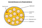
Peripheral Nerve Structure. Sophie Stenner, RVC 2008
Nerve fibres reside in a connective tissue matrix called the endoneurium and are gathered together into bundles or fascicles defined by a second connective tissue layer called the perineurium. Groups of fascicles are then gathered together in a third connective tissue layer called the epineurium. Thus, peripheral nerves have a three-tiered hierarchical arrangement of connective tissue. Renaut bodies are loose, cigar-shaped whorls of extracellular matrix within fascicles that are common in equine nerves at points of stress or compression.
Nerve Fibre
The nerve fibre consists of the impulse-carrying axon, which is surrounded by an ensheathing cell, the Schwann cell, which in turn is surrounded by an acellular basal lamina that is continuous along the length of the nerve. Nerve fibres come in various discrete diameter groups, which are reflected in their conduction velocities. The larger the diameter the more rapid the rate of impulse conduction. Particular targets or receptors are associated with axons of a particular diameter. Axons connected to muscles spindles have a large diameter (20 µm) and conduct at 120 m/s whilst the smallest myelinated fibres are about 1µm and conduct at around 6 m/s. The smallest fibres of all are the unmyelinated fibres (the high-threshold sensory afferents, or C-fibres, and post-ganglionic autonomies) and have a diameter of between 1 and 0.1 µm. These fibres do not conduct by saltatory conduction and have very slow conduction rates of around 0.5 m/s.
Axon
Axons have an outer membrane called the axolemma and within this there is the axoplasm which is continuous with the cytoplasm of the neuron. There are no ribosomes, either free or attached to endoplasmic reticulum in axons and therefore, no protein synthesis. Protein synthesis takes place within the cell body and some dendrites and all protein replacement required for the maintenance of the axon depends on proteins being imported from the cell body. A critical feature of the axon is its cytoskeleton, which consists of two key elements; neurofilaments and microtubules. Neurofilaments are intermediate filaments of about 10 nm diameter, and belong to the same class as other cytoskeletal proteins such as keratin, desmin, vimentin, or GFAP of astrocytes. Neurofilaments are formed from a triplet of polypeptide subunits of heavy (~ 200 kD), medium (~ 150 kD) and low (~ 60 kD) molecular weights. Typically, these subunits are heavily phosphorylated and are more numerous than microtubules, especially in large diameter axons, having a pivotal role in determining axon diameter. They are formed in the cell body, transported down the axon by axoplasmic transport and degraded in the terminals by Ca2+ activated proteases. In other words, there is a constant turnover of neurofilament within the healthy axon. Microtubules within axons are similar to microtubules elsewhere, consisting of polymerised dimers of alpha and beta tubulin arranged as a hollow tube of about 28 nm. They are relatively abundant in smaller diameter axons, and are also synthesised in the cell body. An important component of the cytoskeleton are the microtubule associated proteins or MAP's and the tau proteins. These proteins are important in microtubule assembly and stability. Different classes of MAP's occur in the dendrites and the axons, and to some extent account for the different ultrastructural features that distinguish these two types of neuronal process. They form cross links between adjacent microtubules but also connect to neurofilaments and actin microfilaments, implying complex interactions between the various components of the axon skeleton.
Schwann Cell
Myelination in the PNS is achieved by the Schwann cell, a derivative of neural crest cells, which bud off from the neuroepithelium at a very early stage of neurogenesis. During development, Schwann cells engage many small axons and as axonal diameter increases, Schwann cells eventually relate with only a single axon c.f oligodendrocytes. This single axon is enveloped in a trough by the Schwann cell processes that engulf it and as the processes come together, an inner mesaxon is formed. The leading-edge process continues to move over the axon forming a spiral. Myelination, an extremely complex molecular process, occurs when the cytoplasm within the process is extruded allowing the internal surfaces of the membrane to come together as the major dense line, the outer membrane apposition constituting the intraperiod line. The alternating pattern of these two form the lamellae of compacted myelin. The myelin sheath is attached to, and is an integral part of the Schwann cell on which it is dependent for its maintenance.
A single Schwann cell forms a single myelin sheath or internode and there is a reasonably constant relationship between the myelin thickness and the internodal length, which in turn is associated with axon calibre. Large axons have long, thick myelin sheaths and therefore conduct more rapidly. The internodes do not abut one another but are separated by an exposed area of axon called the node of Ranvier. If the axons remain of small diameter, then a Schwann cell will continue to associate with many axons, although none of them are myelinated. Thus, even unmyelinated axons retain a Schwann cell ensheathment. These non-myelinating Schwann cells are sometimes referred to as Remak cells.
Axoplasmic Transport
Neurons are very large cells and most of a neurons cytoplasm is present in its processes while most of the cells RNA is located in cell body (Nissl substance). These cells have therefore evolved mechanisms to transport large macromolecules and organelles up and down processes.
Anterograde Transport
Anterograde transport moves substances from the cell body to the axon. Two basic forms of anterograde transport can be recognised: fast anterograde transport and slow anterograde transport. Fast anterograde transport allows movement of all membranous organelles such as synaptic vesicles and occurs at a rate of around 400mm/day (recent evidence suggests that there are many forms of fast anterograde transport, mediated by different kinesins). Fast anterograde transport depends critically on oxidative metabolism, and is, in fact independent of the cell body. Microtubules act as a static track along which the organelles can move, driven by the ATPase kinesin which acts as a "motor" molecule. Fast anterograde transport is independent of the cell body. Anything which interferes with energy supply or cytoskeleton necessary for fast anterograde transport has profound effects on the health of the axon. Agents such as colchicine or vincristine block microtubule assembly, disrupting fast anterograde transport. Slow anterograde transport deals with cytoskeletal elements and large soluble proteins. Slow anterograde transport can be further sub-divided into a slow component, which occurs at about 2mm/day (neurofilament, rubulin, actin) and a fast component, which occurs at around 4 mm/day, transporting all other proteins (eg myosin, clathrin).
Retrograde Transport
Retrograde transport returns materials from the axon terminal to the cell body, either for degradation or restoration and reuse. As with fast anterograde transport, particles move along microtubules. The motor molecule for retrograde transport is dynein which is a microtubule-associated ATPase. The retrograde transport system is important not only for returning material to the cell body, but also provides the means whereby target-derived trophic factors, such as nerve growth factor (NGF) for dorsal root ganglion neurons, are conveyed to the cell body where they promote cell survival. Research is being undertaken into the use of trophic factors to promote cell survival during degenerative pathology. The retrograde transport system can be "hijacked" by harmful substances to gain entry to the peripheral neuron and ultimately the CNS. Herpes virus, tetanus and heavy metals all affect the retrograde transport system.
Blood Supply
The epineurium is penetrated by the vascular supply to the nerve and this blood supply is known as the vasa nervorum. Only capillaries occur within the endoneurial compartment. The capillaries of the endoneurium are joined by tight junctions and provide a barrier to large macromolecules. This forms the basis of the blood-nerve barrier (BNB), which has similarities to the blood-brain barrier of the CNS. The BNB appears to be relatively weak in the sensory ganglia because fenestrations occur between endothelial cells in this location. Sensory ganglia are therefore more vulnerable to blood-borne agents. A further "barrier" is provided by the perineurium which consists of sheets of flattened cells, connected by tight junctions and covered on both sides by a basal lamina. The only route across this structure is trans- rather than inter-cellular.
