|
|
| (71 intermediate revisions by 12 users not shown) |
| Line 1: |
Line 1: |
| − | | + | {{toplink |
| | + | |backcolour = FFCCCC |
| | + | |linkpage =Integumentary - Anatomy & Physiology |
| | + | |linktext =INTEGUMENTARY SYSTEM |
| | + | |maplink = Integumentary System (Content Map) - Anatomy & Physiology |
| | + | |pagetype =Anatomy |
| | + | }} |
| | + | <br> |
| | ==Introduction== | | ==Introduction== |
| − | The hoof is defined from a physiologic perspective as the modified skin [[Skin - Anatomy & Physiology#Epidermis|(epidermis)]] covering the tip of the digit and all enclosed structures. The hoof provides protection to the distal limb and is formed by keratinisation of the epithelial layer and modification of the underlying dermis. The keratin in the [[Skin - Anatomy & Physiology#Epidermis|epidermis]], when thickened and cornified, is referred to as [[Horn - Anatomy & Physiology|horn]]. Horn makes up the outer surface if the hoof and is particularly resistant to mechanical and chemical damage.
| + | The keratin in the epidermis, when thickened and cornified, is referred to as [[Horn - Anatomy & Physiology|horn]]. Horn is particularly resistant to mechanical and chemical damage. The outer surface of the hoof is composed of horn. |
| − | | |
| − | Each epidermal region of the hoof is associated with a dermal region (corium). The corium are connected to the underlying structures by the subcutis. As the foot can be divided into 5 segments – Wall, coronary, periople, sole, and frog – there are 5 corresponding underlying corium.
| |
| − | <br />
| |
| − | | |
| − | The origin of the hoof is as a form of protection to the distal phalanx and stems from local modifications of the epidermis, [[Skin - Anatomy & Physiology#Dermis|dermis]] and subcutaneous layers. There is great variation in the shape and types of hooves across different species and this is often due to the multi-role nature of the hoof. In some species the hoof may play an important role in non-locomotion roles such as digging or being used as a weapon. The hoof also acts as a shock-absorber in that the hoof acts to reduce the concussion of foot impact on the structures of the distal limb. It has also been suggested in some species that the elastic nature of components of the hoof may aid the return of blood to the heart. The following information relates primarily to the horse hoof.
| |
| − | | |
| − | ==Five Segments of the Hoof==
| |
| − | ===Wall===
| |
| − | [[File:Lateral_view_horse_foot.jpg|thumb|right|150px|]]
| |
| − | The wall of the hoof is the element of the hoof that is visible. The wall forms the medial, lateral and dorsal aspect of the hoof and it can be further divided into the toe, quarters and heels. At the heel the walls reflect back on themselves at a point called the angles and in doing so forms the bars. The bars, although moving cranially, gradually fade along the edge of the frog and never actually meet.
| |
| − | The wall of the hoof is widest at the distal aspect of the hoof, i.e. at the toe. The wall of the hoof decreases in width laterally and medially (around the quarters of the hoof). In most domestic species the wall of the hoof is between 5 - 10mm thick and consists of three layers. There is an outer layer of thin but dense horn that is shiny and acts to reduce evaporation from the horn and therefore prevent dehydration of the other layers of the horn – the periople. There is an intermediate layer which represents the main structure of the wall and is composed of amorphous horn reinforced with many tubular shaped horn rods. Finally, there is an inner laminar layer where there are interdigitating laminae of horn and dermal laminae which ensure the hoof itself is firmly anchored to the distal phalanx. The bond between the horn and the underlying dermis is a living bond that gradually allows the wall of the hoof to slide in a distal direction towards the ground where the distal border is worn away via contact with the ground. Attached by the subcutis to the periosteum of the distal phalanx, the laminar corium suspends the entire weight of the animal by the distal phalanx within the hoof capsule via microscopic interdigitations with the epidermal laminae of the hoof wall. Damage to the vasculature of the laminar corium can result in compromises in the integrity of the interdigitations.
| |
| − | | |
| − | The hooves in newborn foals are bilaterally symmetrical. Over a period of just a few months, forces exerted on the hoof during locomotion cause a visible difference between the right and left, as well as front and hind hooves. Thus, isolated specimens of equine feet can be distinguished as follows:
| |
| − | | |
| − | Front vs hind:
| |
| − | Front: The angle between the toe and the ground is approximately 45 degrees. The sole is circular in shape.
| |
| − | Hind: The angle between the toe and the ground is 50-55 degrees. The sole is oval in shape.
| |
| | | | |
| − | Right vs left:
| + | ==Structure and Function== |
| − | Quarters (lateral and medial walls) are steeper on the medial side of the hoof.
| |
| − | Assessment of the external anatomy can be a used as an important part of a lameness examination.
| |
| | | | |
| − | ===Periople=== | + | ===Equine Hoof=== |
| − | The periople is a small band of soft tissue found over the proximal surface of the wall of the hoof. The periople represents the junction between the wall and the skin of the limb. The periople is responsible for the production of the outer layer of the wall (above). The periople segment extends around the proximal edge of the wall and in horses covers the bulb of the heel and part of the frog. In ruminants it forms part of the join between both hooves of the foot. The perioplic corium, proximal to the coronary band and continuous with the skin, it produces the thin, shiny layer (stratum externa) of the hoof wall.
| |
| | | | |
| | + | The epidermis of the horny hoof is termed the '''coronary epidermis'''. Its surface has a plantar aspect and newly formed hoof grows distally towards the plantar surface of the structure. Hoof wall is generally 5 - 10 mm in thickness and consists of 3 layers: |
| | + | * A thin outer layer or "'''wall'''" of shiny, dense horn, which effectively seals the hoof against dehydration and penetration. |
| | + | * A thicker, intermediate layer of '''amorphorous''' horn, interspersed with '''tubular horn''' which provides reinforcement. This layer makes up the bulk of the hoof. |
| | + | * An inner, lamellar layer, where epidermis and dermis interdigitate, anchoring the dead portion of the hoof to the living surface of the third phalanx. |
| | | | |
| − | ===Coronary Segment===
| |
| | [[image: X-section through hoof.jpg|thumb|200px|right|A X-section through a typical hoof. ©Rachael Wallace2008]] | | [[image: X-section through hoof.jpg|thumb|200px|right|A X-section through a typical hoof. ©Rachael Wallace2008]] |
| − | The wall of the hoof is created at the coronary dermis and grows in a distal direction from the coronary dermis. The coronary dermis is studded with many papillae which are directed towards the ground in the direction of growth. The epidermis covering these papillae produce horn tubules which are embedded into amorphous inter-tubular horn. This inter-tubular horn is created by the spaces between the papillae within the coronary dermis. The combination of both of these horn types ensures the horn has sufficient strength.
| |
| − |
| |
| − | The pigmentation of the hoof is derived from melanocytes found in the coronary epidermis. Any pigmentation in the hoof will be most pronounced in the outer part of the hoof wall as the deeper layers of the hoof usually contain fewer melanocytes. It is this unpigmented element of the hoof that forms the 'white line' in the sole of hoofs and is particularly important in horses as a landmark for shoeing.
| |
| | | | |
| − | The coronary corium is responsible for the growth of the bulk of the tubular and non-tubular horn that make up the hoof wall. This wall glides distally at a rate of 5-6mm a month and by forming epidermal laminae itself it interdigitates with the underlying dermal laminae. Neither of these laminae are pigmented so when the epidermal laminae appear on the solar surface, a non-pigmented region known as the white line appears. The white line is used as important landmark in farriery as structures central to the line will be dermal and so vascular and sensitive. Interruptions in the coronary corium can result in defective hoof wall growth.
| |
| | | | |
| − | ===Sole Segment===
| + | The pigmentation in the outer layer of hooves is derived from melanocytes in the coronary epidermis. Deeper layers contain little melanin and therefore appear lightly coloured or white. The unpigmented layer of keratin forms a '''white line''' on the sole of the hoof, which delineates the sole from the wall when the plantar aspect is viewed. |
| − | [[image: Plantar hoof aspect.jpg|thumb|175px|left|A view of the solar surface of an equine hoof. The wall has been removed on the right to show the underlying dermis. ©Rachael Wallace2008]]
| |
| − | The sole is the area distal to the bars and apex of the frog enclosed by the hoof wall. The area where the bars and wall enclose it is known as the angle of the sole. Since the sole is slightly concave, the majority of the horse's weight is transferred through the margin of the sole.
| |
| | | | |
| − | The sole represents the part of the foot in contact with the ground and its composition differs between species. The keratin found in the sole is formed from the epidermis on the underside of the third phalanx and can grow to a thickness of around 10mm in domestic species. The keratin found on the sole is much more easily worn down or abraded than that of the wall of the hoof. The equine sole has a central frog structure whilst ruminants and pigs have a bulb structure to the sole.
| + | [[image: Plantar hoof aspect.jpg|thumb|175px|left|A view of the plantar aspect of an equine hoof. The wall has been removed on the right to show the underlying dermis. ©Rachael Wallace2008]] |
| | | | |
| − | The solar corium is the dermal layer underlying the solar surface that produces a superficially flaky epidermis. Sufficient solar depth is necessary to protect the underlying soft tissue and bony structures. | + | The keratin in the sole is formed by the epidermis of the plantar aspect of the third phalanx and reaches a maximum thickness of 1 cm. It is less resistant to abrasion than that of the outer layer of the hoof. The central '''frog''' of the sole of a horse is located between the sharply inflected '''bars''' of the wall and is softer and less rigid than the keratin of the wall of the hoof. Its elasticity is particulary important in the horse as pressure on the frog changes the angle of the walls when the horse stands on that foot. The change in angle, termed the '''hoof mechanism''' increases the blood circulation in the hoof, increasing the supply of nutrients to the coronary epidermis. This is closely correlated to hoof wall quality. |
| | | | |
| − | ===Frog-Bulb Segment===
| + | A band of soft, pliable horn termed the '''periople''' (analagous with the cuticle of human nails) lies over the proximal outer surface of the wall. This band widens at the back of the hoof to also cover the '''bulbs of the heels''' and part of the '''frog'''. |
| − | The frog is a wedge-shaped structure which sits between the bars and has an apex facing distally, with 2 crura flanking a central sulcus. Between the crus and bar of each half of the sole lies the collateral sulcus. Opposite the apex, the frog expands forming the bulbs of the heel. The frog is a mass of keratinized stratified squamous epithelium, which is softer than other parts of the hoof due to its increased water content. Usually, the frog contributes to the weightbearing surface where it functions as a shock absorber. Apocrine glands within the corium of the frog produce secretions on the surface. The frog ensures that the wall of the hoof is forced outwards when weight is put on the limb thus ensuring that the 'hoof mechanism' functions correctly and ensuring circulatory flow around the hoof and back towards the heart.
| |
| − | The ruminant/pig 'bulb' provides the hoof with the caudal and mid-hoof contact area with the ground and is chiefly involved in weight bearing. The bulb inserts into the V-shaped sole. The bulb is made of relatively soft material, mainly inter-tubular horn and is of a considerable thickness.
| |
| | | | |
| − | The frog corium overlies the digital cushion and generates the specialised soft epidermal tissues of the frog.
| + | ===Ruminant Hoof=== |
| − | | |
| − | ==Deeper structures of the foot== | |
| − | [[File:Horse_hoof_wild_bare_sagittal.jpg|thumb|200px|right|Sagittal section through horse hoof.]]
| |
| − | Enclosed within the hoof capsule are the bony structures – the [[Phalanges_-_Horse_Anatomy#Distal_Phalanx|distal phalanx]], distal end of the [[Phalanges_-_Horse_Anatomy#Middle_Phalanx|middle phalanx]], the [[Joints_and_Ligaments_-_Horse_Anatomy#Distal_Interphalangeal_.28Coffin.29_Joint|distal interphalangeal joint]], and the [[Phalanges_-_Horse_Anatomy#Distal_Sesamoid_.28Navicular.29_Bone|distal sesamoid bone (navicular)]]. There are also soft tissue structures including ligaments, cartilage, the digital cushion and the insertions of the [[Tendons_-_Horse_Anatomy#Thoracic_Limb|common digital extensor tendon]] and the [[Tendons_-_Horse_Anatomy#Flexorsdeep|digital flexor tendon]].
| |
| − | | |
| − | ===Ungual (collateral) cartilages=== | |
| − | The ungual cartilages are extensions of the [[Phalanges_-_Horse_Anatomy#Distal_Phalanx|distal phalanx]] (Plll) that extend caudally and dorsally from the medial and lateral margins of the [[Phalanges_-_Horse_Anatomy#Distal_Phalanx|distal phalanx]], curving inwards towards each other in the heel region. The cartilages extend just beyond the confines of the hoof capsule making them palpable just above the coronary band at the lateral and medial edges of the foot. The ungual cartilages can ossify resulting in ‘side bones’ which have the potential for fracturing. The cartilages can also become infected resulting in the condition known as ‘quittor’.
| |
| − | | |
| − | The cartilages are securely attached to the other internal structures of the foot by a series of ligaments that extend from the medial and lateral cartilages to the distal and middle phalanx, the [[Phalanges_-_Horse_Anatomy#Distal_Sesamoid_.28Navicular.29_Bone|distal sesamoid bone (navicular)]], and the digital cushion.
| |
| − | | |
| − | [[File:Lateral view hoof internal structures.jpg|thumb|Internal hoof structures]]
| |
| − | | |
| − | ===Collateral ligaments.===
| |
| − | The distal interphalangeal joint is enclosed within the hoof capsule. It is stabilised by the medial and lateral collateral ligaments which form part of the joint capsule, connecting the distal end of Pll with the proximal edge of the [[Phalanges_-_Horse_Anatomy#Distal_Phalanx|distal phalanx]].
| |
| − | | |
| − | [[File:Oblique view.jpg|thumb|Internal hoof structures]]
| |
| − | | |
| − | ===Annular ligaments===
| |
| − | The annular ligament has its origins on the medial and lateral surfaces of distal Pl. It is the most superficial structure in the region, lying just beneath the skin and fusing with the [[Tendons_-_Horse_Anatomy#Flexorsdeep|digital flexor tendon]] where it enters the hoof capsule. Once inside the hoof the annular ligament merges with the fibrous attachments of the ungual cartilages and digital cushion, and continues with the [[Tendons_-_Horse_Anatomy#Flexorsdeep|digital flexor tendon]] down to its insertion onto the [[Phalanges_-_Horse_Anatomy#Distal_Phalanx|distal phalanx]].
| |
| − | | |
| − | ===Sesamoidean ligaments===
| |
| − | The dorsal border of the [[Phalanges_-_Horse_Anatomy#Distal_Sesamoid_.28Navicular.29_Bone|distal sesamoid bone (navicular)]] is held securely to the palmar/plantar surface of the [[Phalanges_-_Horse_Anatomy#Distal_Phalanx|distal phalanx]] by the distal sesamoidean ligament and to the proximal phalanx via the proximal interphalangeal collateral ligaments by means of a pair of medial and lateral collateral sesamoidean ligaments.
| |
| − | | |
| − | ===Navicular bursa===
| |
| − | The [[Phalanges_-_Horse_Anatomy#Distal_Sesamoid_.28Navicular.29_Bone|distal sesamoid bone (navicular)]] lies between the middle and distal phalanges and the deep digital flexor tendon. Associated with it is a fluid-filled sac that reduces friction between the bone and the [[Tendons_-_Horse_Anatomy#Flexorsdeep|digital flexor tendon]] that lies over the top of it— the navicular bursa. Inflammation in the region is involved in navicular disease which is a common cause of lameness.
| |
| − | | |
| − | | |
| − | ===Digital Cushion===
| |
| − | The digital cushion is the internal tissue deep to the frog. It lies between the ungual cartilages and is comprised of collagenous, elastic tissue infiltrated by adipose tissue. At the bulbs of the heel, it is subcutaneous and is soft and loose in texture. It has connection with the digital annular ligament and, at the apex to the deep digital flexor tendon at its point of insertion on the distal phalanx. It acts as one of the major shock absorbers of the foot. When the limb is weight bearing, the increase in pressure and change in shape of the digital cushion and the frog compress the veins in the foot aiding venous return.
| |
| − | | |
| − | ===Blood supply to the digit===
| |
| − | The main vessels supplying the digit in the forelimb are the medial and lateral palmar digital aa, both of which arise from the median a. In the digit of the hind limb the medial and lateral digital aa. are a continuation of the metatarsal a. and are also contributed to by the medial and lateral plantar aa. which branch from the sapheneous a.
| |
| − | The digital arteries give rise to numerous branches forming rich networks for the vascular tissues. Many anastomoses occur. The terminal branches of the main vessels finally enter a bony canal in the distal phalanx. Venous drainage is similar with the most distal vessels being the medial and lateral palmar/plantar digital veins. The compressive action of the hoof on the soft tissues within during locomotion generates an important function promoting venous return.
| |
| − | | |
| − | Normal equine digital vasculature anatomy can be divided into five major areas of perfusion:
| |
| − | 1. Coronary plexus
| |
| − | 2. Dorsal lamellar plexus
| |
| − | 3. Circumflex vessels
| |
| − | 4. Terminal arch
| |
| − | 5. Heel perfusion.
| |
| − | Loss of perfusion to the lamella vessels, circumflex vessels, and terminal arch indicates a poor prognosis without aggressive therapy.
| |
| − | | |
| − | ===Microcirculation in the dermal laminae===
| |
| − | Numerous arteriovenous anastomoses occur which are of a somewhat unusual type. Under normal circumstances these are closed and as a result circulation within the capillary beds of the dermal laminae occurs. Certain systemic pathologies may result in opening of these AV anastomoses resulting in ischaemia of the laminae. This in turn results in the hoof wall separating from the distal phalanx producing the disease termed “laminitis”, which can be either acute or chronic.
| |
| − | | |
| − | ===Innervation of the equine digit===
| |
| − | The digit of the forelimb is innervated by the medial and lateral digital nerves. The medial digital n. is a continuation of the median n. and the lateral digital n. is derived from both median and ulnar nerves. These run on the palmar aspect of the digit in close proximity to the main arteries and veins. They give rise to several dorsal branches which supply dorsally located areas.
| |
| | | | |
| − | The pelvic limb digit is innervated on the dorsal aspect by the common digital nn. Derived from the fibular n. The plantar aspect is innervated by the medial and lateral digital nn which originate from the tibial n.
| + | Although the hooves of these species resemble the equine hoof, they differ in several ways: |
| − | In addition to their normal importance in supplying innervation to the sensitive tissues of the equine digit these nerves are also of considerable clinical importance as they are utilized for the procedure termed diagnostic nerve blocks.
| |
| | | | |
| − | ==Species variation==
| + | * There are two separate main digits, compared with the single hoof of equids |
| | + | * The wall is bent to form a dorsal border |
| | + | * The bulb of the heel covers the entire caudal surface of the hoof and most of the plantar surface, leaving a small area of sole visible |
| | + | * The interdigitating lamellae are smaller and less well developed. |
| | | | |
| − | ===Ruminant Hoof===
| + | The hooves of the main digits curve medially towards each other. The lateral digit carries more weight than the medial digit, and is larger. On the abaxial wall, the distal border makes contact with the ground along its entire length, whereas, on the axial wall, only does so toward the toe. The thickness of the wall increases towards the apex and the plantar surface. |
| − | The ruminant hoof, although resembling the equine hoof in some characteristics, differs from the equine hoof in several ways. In the ruminant hoof there are two separate main digits and the wall of the hoof is bent to form a border. Also the bulb of the heel covers the entire caudal surface of the hoof and most of the plantar surface, leaving only a small area of sole visible. In ruminants the interdigitating lamellae are smaller and less well developed than in equids.
| |
| − | <br />
| |
| − | <br />
| |
| − | The hooves of the main digits curve medially towards each other. The lateral digit carries more weight than the medial digit, and is larger. On the abaxial wall, the distal border makes contact with the ground along its entire length, whereas, on the axial wall, only does so toward the toe. The thickness of the wall increases towards the apex and the plantar surface. The horn of the hoof generally grows at a rate of 5 mm per month, and in cattle allowed to move freely, growth should equal wear. In intensively kept cattle, growth exceeds wear, and foot trimming is required to maintain optimal shape and angle. The optimal angle of the toe from the ground is 50 degrees. Where horn overgrowth occurs, the coffin joint is gradually overextended and the deep flexor tendon tensed. This results in greater weight being placed over the caudal part of the hoof and can cause pain and lameness. | |
| − | <br />
| |
| − | <br />
| |
| − | '''Dewclaws''' are present in most ruminants but do not make contact with the ground. They consist of wall and bulb and have no practical importance.
| |
| | | | |
| | + | The horn of the hoof generally grows at a rate of 5 mm per month, and cattle allowed to move freely, growth should equal wear. In intensively kept cattle, growth exceeds wear, and foot trimming is required to maintain optimal shape and angle. The optimal angle of the toe from the ground is 50 degrees. |
| | | | |
| − | See the [[Bovine Lower Limb - Anatomy & Physiology|bovine lower limb]] for further detail.
| + | '''Dewclaws''' are present in most ruminants but do not make contact with the ground. They consist of wall and bulb and have no practical importance. |
| | | | |
| | ===Porcine Hoof=== | | ===Porcine Hoof=== |
| − | [[File:Pig cracked hooves.JPG|thumb|right|150px|Pig Hooves and a cracked hoof wall]]
| + | The hooves of pigs are principally similar to those of ruminants, however the wall is straight, not bent medially at the toe, and have a soft bulb that is well distanced from the wall and sole. The hooves of the accessory digits are of the same structure as the principal digits, but only bear weight on soft ground. |
| − | The hooves of pigs are principally similar to those of ruminants, however the wall is straight, not bent medially at the toe, and they have a soft bulb that is well distanced from the wall and sole. The hooves of the accessory digits are of the same structure as the principal digits, but only bear weight on soft ground. | |
| | | | |
| | Hoof trimming in pigs is rarely required due to the short lifespan of the farmed pig. | | Hoof trimming in pigs is rarely required due to the short lifespan of the farmed pig. |
| − |
| |
| − | {{Learning
| |
| − | |flashcards = [[Hoof flashcards - Anatomy & Physiology|Hoof Flashcards]]
| |
| − | |full text = [http://www.cabi.org/cabdirect/FullTextPDF/2005/20053192986.pdf ''' The growth and adaptive capabilities of the hoof wall and sole: functional changes in response to stress.''' Bowker, R. M.; American Association of Equine Practitioners (AAEP), Lexington, USA, Proceedings of the 49th Annual Convention of the American Association of Equine Practitioners, New Orleans, Louisiana, USA, 21-25 November 2003, 2003, pp 146-168, 32 ref.]<br>[http://www.cabi.org/cabdirect/FullTextPDF/2005/20053192988.pdf '''Contrasting structural morphologies of "good" and "bad" footed horses.''' Bowker, R. M.; American Association of Equine Practitioners (AAEP), Lexington, USA, Proceedings of the 49th Annual Convention of the American Association of Equine Practitioners, New Orleans, Louisiana, USA, 21-25 November 2003, 2003, pp 186-209, 73 ref.]
| |
| − | |OVAM = [http://www.onlineveterinaryanatomy.net/content/white-line-bovine-hoof White Line of Bovine Hoof]<br>[http://www.onlineveterinaryanatomy.net/content/hoof-wall-bovine-distal-limb Bovine Hoof Wall]
| |
| − | }}
| |
| − |
| |
| − |
| |
| − | ==Webinars==
| |
| − | <rss max="10" highlight="none">https://www.thewebinarvet.com/herd-health/webinars/feed</rss>
| |
| − | {{review}}
| |
| − | [[Category:Integumentary System - Anatomy & Physiology]]
| |
| − | [[Category:A&P Done]]
| |
Introduction
The keratin in the epidermis, when thickened and cornified, is referred to as horn. Horn is particularly resistant to mechanical and chemical damage. The outer surface of the hoof is composed of horn.
Structure and Function
Equine Hoof
The epidermis of the horny hoof is termed the coronary epidermis. Its surface has a plantar aspect and newly formed hoof grows distally towards the plantar surface of the structure. Hoof wall is generally 5 - 10 mm in thickness and consists of 3 layers:
- A thin outer layer or "wall" of shiny, dense horn, which effectively seals the hoof against dehydration and penetration.
- A thicker, intermediate layer of amorphorous horn, interspersed with tubular horn which provides reinforcement. This layer makes up the bulk of the hoof.
- An inner, lamellar layer, where epidermis and dermis interdigitate, anchoring the dead portion of the hoof to the living surface of the third phalanx.
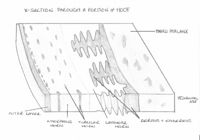
A X-section through a typical hoof. ©Rachael Wallace2008
The pigmentation in the outer layer of hooves is derived from melanocytes in the coronary epidermis. Deeper layers contain little melanin and therefore appear lightly coloured or white. The unpigmented layer of keratin forms a white line on the sole of the hoof, which delineates the sole from the wall when the plantar aspect is viewed.
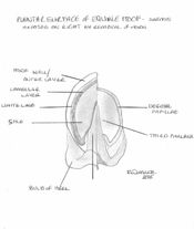
A view of the plantar aspect of an equine hoof. The wall has been removed on the right to show the underlying dermis. ©Rachael Wallace2008
The keratin in the sole is formed by the epidermis of the plantar aspect of the third phalanx and reaches a maximum thickness of 1 cm. It is less resistant to abrasion than that of the outer layer of the hoof. The central frog of the sole of a horse is located between the sharply inflected bars of the wall and is softer and less rigid than the keratin of the wall of the hoof. Its elasticity is particulary important in the horse as pressure on the frog changes the angle of the walls when the horse stands on that foot. The change in angle, termed the hoof mechanism increases the blood circulation in the hoof, increasing the supply of nutrients to the coronary epidermis. This is closely correlated to hoof wall quality.
A band of soft, pliable horn termed the periople (analagous with the cuticle of human nails) lies over the proximal outer surface of the wall. This band widens at the back of the hoof to also cover the bulbs of the heels and part of the frog.
Ruminant Hoof
Although the hooves of these species resemble the equine hoof, they differ in several ways:
- There are two separate main digits, compared with the single hoof of equids
- The wall is bent to form a dorsal border
- The bulb of the heel covers the entire caudal surface of the hoof and most of the plantar surface, leaving a small area of sole visible
- The interdigitating lamellae are smaller and less well developed.
The hooves of the main digits curve medially towards each other. The lateral digit carries more weight than the medial digit, and is larger. On the abaxial wall, the distal border makes contact with the ground along its entire length, whereas, on the axial wall, only does so toward the toe. The thickness of the wall increases towards the apex and the plantar surface.
The horn of the hoof generally grows at a rate of 5 mm per month, and cattle allowed to move freely, growth should equal wear. In intensively kept cattle, growth exceeds wear, and foot trimming is required to maintain optimal shape and angle. The optimal angle of the toe from the ground is 50 degrees.
Dewclaws are present in most ruminants but do not make contact with the ground. They consist of wall and bulb and have no practical importance.
Porcine Hoof
The hooves of pigs are principally similar to those of ruminants, however the wall is straight, not bent medially at the toe, and have a soft bulb that is well distanced from the wall and sole. The hooves of the accessory digits are of the same structure as the principal digits, but only bear weight on soft ground.
Hoof trimming in pigs is rarely required due to the short lifespan of the farmed pig.

