Lingual Gland - Anatomy & Physiology
Jump to navigation
Jump to search
Overview

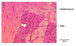
Serous Lingual Gland Histology (Mouse), from oral cavity tutorial part 1, slide 31

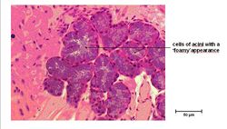
Mucous Lingual Gland Histology (Mouse), from oral cavity tutorial part 1, slide 33
Acini with cuboidal epithelium, round basal nuclei and cytoplasm that stains pink is a serous secreting lingual gland in the body of the tongue.
Acini with flattened basal nuclei, cytoplasm that stains blue and has a foamy appearence is a mucous secreting lingual gland in the root of the tongue.
| Lingual Gland - Anatomy & Physiology Learning Resources | |
|---|---|
To reach the Vetstream content, please select |
Canis, Felis, Lapis or Equis |
 Selection of relevant PowerPoint tutorials |
Oral Cavity part 1 tutorial covers lingual glands |
Error in widget FBRecommend: unable to write file /var/www/wikivet.net/extensions/Widgets/compiled_templates/wrt662a1243aa6c62_11101137 Error in widget google+: unable to write file /var/www/wikivet.net/extensions/Widgets/compiled_templates/wrt662a1243ad86d8_54090809 Error in widget TwitterTweet: unable to write file /var/www/wikivet.net/extensions/Widgets/compiled_templates/wrt662a1243b15257_28262225
|
| WikiVet® Introduction - Help WikiVet - Report a Problem |