Meninges - Anatomy & Physiology
Jump to navigation
Jump to search
| This article is still under construction. |

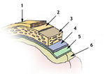
Diagram of the meninges. Sourced from WikiMedia Commons, attributed to http://training.seer.cancer.gov/module_anatomy/unit5_3_nerve_org1_cns.html
- The CNS is surrounded by several layers of tissue.
- Bone layer with periosteum
- E.g. the skull and the vertebrae.
- Epidural Space
- Lies between periosteum and dura in vertebral canal. Contains loose connective tissue, veins and lymphatics. Cushions the cord as it flexes. Can be used for nerve blocks (epidural). In cranium, dura fused with periosteum, one single layer in effect.
- Dura mater
- dense connective tissue
- fused with the periosteum of cranium
- contains the venous sinuses.
- Folds of dura mater
- Falx cerebri- midline fold between cerebral hemispheres
- Tentorium cerebelli- oblique fold between the cerebrum and cerebellum
- Diaphragma sellae- forms a collar around the neck of the pituitary, forms the roof of the hypophyseal fossa
- Subdural space
- Lies between dura and arachnoid (not fused). Potential space containing only lymph-like fluid. Site of subdural hematoma.
- Arachnoid mater
- Delicate, non-vascular connective tissue
- Adhered to the skull in the calvaria.
- Subarachnoid space
- between arachnoid and pia connected by tiny filaments (spider-like)
- contains CSF from ventricular system
- largest parts are the cisterns
- Cerebellomedullary cistern around the foramen magnum. Used for the collection of CSF
- Lumbar cistern used for lumbar puncture in man
- Pia mater
- highly vascular connective tissue, very closely applied to brain tissue
- Bone layer with periosteum
- The epidural space lies between the bone layer (skull or vertebrae) and the dura-arachnoid layer.
- Local anaesthetic is injected here in epidural anaesthesia.
- E.g. when calving cows.
- Local anaesthetic is injected here in epidural anaesthesia.
- The sub-arachnoid space lies between the arachnoid layer and the pia mater.
- CSF filled.
- This is where myelographic contrast medium is injected.