Meninges - Anatomy & Physiology
Outer Layers
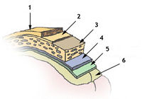
The central nervous system (CNS) is surrounded by several layers of tissue, with several outer layers not directly related to the CNS and three membranes that directly envelope the CNS. The outer layers are the skin and then a bone layer with associated periosteum. This layer includes the skull and the vertebrae. Below the periosteum is the Epidural Space which lies between periosteum and dura in the vertebral canal. The epidural space contains adipose tissue, loose connective tissue, veins and lymphatics. It cushions the cord as it flexes. The epidural space is regularly used for nerve blocks where a local anaesthetic such as Lidocaine is injected to block signal transmission to and from the spinal cord.
Dura mater
The Dura mater is the outer menix and is made up of a dense fibrous connective tissue. In the cranium, the dura layer is fused with the periosteum and therefore is in effect single layer without an epidural space. The dura contains a number of folds throughout its coverage of the brain including the Falx cerebri, a midline fold between cerebral hemispheres, the Tentorium cerebelli, an oblique fold between the cerebrum and cerebellum and the Diaphragma sellae which forms a collar around the neck of the pituitary and forms the roof of the hypophyseal fossa. This layer and these associated folds all provide structural support to the brain and prevent the brain from undergoing excess movement within the skull. Where the dura mater folds between brain tissues it splits into two distinct layers that are separated by large blood filled spaces called venous sinuses. Venous sinuses are directly connected to the venous system and venous blood from vessels supplying the brain return to the heart via these sinuses.
Subdural space
The subdural space lies between the dura and the next meningial layer, the arachnoid mater (not fused). The subdural space is thought to contain only lymph-like fluid. This space can also be the site of a subdural hematoma.
Arachnoid mater
This is the middle meningial layer and lies between the dura mater and the pia mater, the innermost meningeal layer. The arachnoid mater is a delicate structure and is constructed with non-vascular connective tissue. This layer also has small protrusions through the dura mater into the previously mentioned venous sinuses called Arachnoid villus and these allow cerebrospinal fluid (CSF) to enter and exit the blood stream. These protrusions adhere to the inner surface of the skull via calvaria processes.
Subarachnoid Space
The subarachnoid space lies between the arachnoid mater and pia mater. Both meninges are connected via a fine network of connective tissue filaments (spider-like) which run through the space, originating from the arachnoid mater. This space also contains CSF from ventricular system. The largest part of this space are called the cisterns which are used for the collection of CSF. For example there is a cerebellomedullary cistern around the foramen magnum. The lumbar cistern used for lumbar puncture in man.
Pia Mater
This is the innermost layer and is firmly bound to the underlying neural tissue of the brain and spinal cord. The inner surface of the brain facing this meningial layer is lined with ependymal cells. The pia mater is highly vascular and is formed from connective tissue.
Error in widget FBRecommend: unable to write file /var/www/wikivet.net/extensions/Widgets/compiled_templates/wrt69a0693e1c8aa5_84605856 Error in widget google+: unable to write file /var/www/wikivet.net/extensions/Widgets/compiled_templates/wrt69a0693e210444_97816513 Error in widget TwitterTweet: unable to write file /var/www/wikivet.net/extensions/Widgets/compiled_templates/wrt69a0693e25a3f8_15483322
|
| WikiVet® Introduction - Help WikiVet - Report a Problem |
