Cerebral Spinal Fluid - Anatomy & Physiology
Cerebrospinal Fluid Function
Cerebrospinal fluid (CSF) surrounds the brain as well as the central canal of the spinal cord. It helps cushion the central nervous system (CNS), acting in a similar manner to a shock absorber. It also acts as a chemical buffer providing immunological protection and a transport system for waste products and nutrients. The CSF also provides buoyancy to the soft neural tissues which effectively allows the neural tissue to "float" in the CSF. This prevents the brain tissue from becoming deformed under its own weight. It acts as a diffusion medium for the transport of neurotransmitters and neuroendocrine substances.
CSF Production and Constituents
CSF is a clear fluid produced by dialysis of blood in the choroid plexus. Choroid plexi are found in each lateral ventricle and a pair are in the third and fourth ventricle. Further production also comes from the ependymal cell linings and vessels within the pia mater.
Ependymal cell production of CSF is via ultrafiltration of blood plasma and active transport across the ependymal cells. The ependyma is connected via a series of tight junctions preventing molecules passing between cells. The ependyma also sits on a basement membrane to provide support to the ependymal cells and provide further protection against blood perfusion. In areas of the brain where there are choroid plexi, the endothelium of the plexus vessel sits immediately adjacent to the basement membrane of the ependymal cells. Of the total CSF production, 35% is produced within the third ventricle of the brain, 23% via the fourth ventricle and 42% from general ependymal cell filtration.
CSF has a very low protein constituent, with only albumin being present together with a very low level of cellularity. The biochemistry of CSF includes high concentrations of sodium and chloride and very high concentrations of magnesium. Concentrations of potassium, calcium and glucose are low.
CSF Circulation
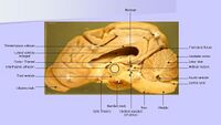
Once produced, CSF is then circulated, due to hydrostatic pressure, from the choroid plexus of the lateral ventricles, through the interventricular foramina into the 3rd ventricle. The lateral ventricles are paired and are located in the cerebral hemispheres. The 3rd ventricle is located in the diencephalon and surrounds the thalamus. CSF then flows through the cerebral aqueduct (aqueduct of Sylvius or mesencephalic aqueduct) into the 4th ventricle. The 4th ventricle is located in the hindbrain. From the 4th ventricle the CSF may flow down the central canal of the spinal cord, or circulate in the subarachnoid space. The central canal of the spinal cord is in direct communication with the 4th ventricle. Most CSF escapes from the ventricular system at the hindbrain Foramen of Luschka (lateral apertures) into the subarachnoid space. Once in the subarachnoid space, the CSF may enter the cerebromedullary cistern (a dilation of the subarachnoid space between the cerebellum and the medulla) and then circulate over the cerebral hemispheres. CSF also flows down the length of the spinal cord in the subarachnoid space. Another dilation of the subarachnoid space occurs caudally due to the dura and arachnoid meninges continuing on past the end of the spinal cord. This gives rise to the Lumbar cistern, and can be used to take a CSF sample in large species. In some species there is an exit which allows CSF to flow from the caudal end of the central canal into the lumbar cistern.
Large amounts of CSF are drained into venous sinuses through arachnoid granulations in the dorsal sagittal sinus. The dorsal sagittal sinus is located between the folds of dura, known as the falx cerebri, covering each of the cerebral hemispheres. Arachnoid granulations contain many villi that are able to act as a one way valve helping to regulate pressure within the CSF, and these arachnoid villi push through the dura and into the venous sinuses.
Because CSF is continuously produced, any disruption to this flow can result in increased pressure building up within parts of the CSF system which can cause compression of neural structures surrounding the area of increased pressure. Clinical examples of this are hydrocephalus and syringohydromyelia.
| This article has been peer reviewed but is awaiting expert review. If you would like to help with this, please see more information about expert reviewing. |
Error in widget FBRecommend: unable to write file /var/www/wikivet.net/extensions/Widgets/compiled_templates/wrt698774f7eb08d3_45026661 Error in widget google+: unable to write file /var/www/wikivet.net/extensions/Widgets/compiled_templates/wrt698774f7f0a1a7_29996902 Error in widget TwitterTweet: unable to write file /var/www/wikivet.net/extensions/Widgets/compiled_templates/wrt698774f805d4a7_32852010
|
| WikiVet® Introduction - Help WikiVet - Report a Problem |
