Difference between revisions of "Mucous Salivary Gland - Anatomy & Physiology"
| (5 intermediate revisions by 2 users not shown) | |||
| Line 1: | Line 1: | ||
| − | + | {{OpenPagesTop}} | |
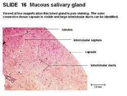
[[Image:Mucous Salivary Gland.jpg|thumb|right|250px|Mucous Salivary Gland Histology - Copyright RVC 2008]] | [[Image:Mucous Salivary Gland.jpg|thumb|right|250px|Mucous Salivary Gland Histology - Copyright RVC 2008]] | ||
==Overview== | ==Overview== | ||
| − | The '''mucous salivary gland''' | + | The '''mucous salivary gland''' is pale staining and is lobed. It has large, interlobular ducts in a connective tissue septum. It has an outer connective tissue capsule. The mucous acini produce a mucous secretion which is a viscous mix of glycoproteins. The cuboidal cells are filled with mucous droplets giving a ‘foamy’ appearance. The nucleus is displaced and flattened near the base of the cell. The mucous cells only stain faintly. |
[[Category:Salivary Glands - Anatomy & Physiology]] | [[Category:Salivary Glands - Anatomy & Physiology]] | ||
| − | [[Category: | + | [[Category:A&P Done]] |
| − | [[ | + | |
| + | {{Template:Learning | ||
| + | |powerpoints = [[Oral Cavity Histology resource|Oral cavity histology tutorial that looks at salivary glands]] | ||
| + | |Vetstream = [https://www.vetstream.com/canis/search?s=salivary Salivary Gland Diseases] | ||
| + | }} | ||
| + | |||
| + | {{OpenPages}} | ||
Latest revision as of 10:06, 7 May 2016
Overview
The mucous salivary gland is pale staining and is lobed. It has large, interlobular ducts in a connective tissue septum. It has an outer connective tissue capsule. The mucous acini produce a mucous secretion which is a viscous mix of glycoproteins. The cuboidal cells are filled with mucous droplets giving a ‘foamy’ appearance. The nucleus is displaced and flattened near the base of the cell. The mucous cells only stain faintly.
| Mucous Salivary Gland - Anatomy & Physiology Learning Resources | |
|---|---|
To reach the Vetstream content, please select |
Canis, Felis, Lapis or Equis |
 Selection of relevant PowerPoint tutorials |
Oral cavity histology tutorial that looks at salivary glands |
Error in widget FBRecommend: unable to write file /var/www/wikivet.net/extensions/Widgets/compiled_templates/wrt6648cf7f28a789_90449671 Error in widget google+: unable to write file /var/www/wikivet.net/extensions/Widgets/compiled_templates/wrt6648cf7f2c1375_19692240 Error in widget TwitterTweet: unable to write file /var/www/wikivet.net/extensions/Widgets/compiled_templates/wrt6648cf7f2f4b13_91483861
|
| WikiVet® Introduction - Help WikiVet - Report a Problem |
