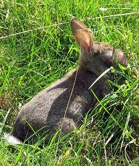
Myxomatosis
Also known as Rabbit Pox
Introduction
Myxomatosis is a highly contagious viral condition of rabbits caused by the myxoma virus, a member of the poxvirus group. It was first recognised in the UK in 1953 after it crossed the channel from France where it was illegally introduced in 1952. It is carried mainly by arthropods, particularly the rabbit flea, Spilopsyllus cuniculi. The disease is also transmitted by direct or indirect contact with ocular or skin discharges or by mechanical vectors. The disease is characterised by subcutaneous mucinous lesions and nodular tumours and is associated with a high mortality rate.
Myxomatosis is enzootic in cottontail rabbits of the genus Sylvilagus in both South and North America and in wild rabbits of the genus Oryctolagus in South America, Europe, and Australia. All other animals are resistant to the disease.
Pathogenesis
The myxoma virus infects several cell types including mucosal cells, lymphocytes and fibroblasts. In addition to primary and secondary tumour development, there is severe immunosuppression leading to overwhelming infections by opportunistic gram-negative bacteria particularly affecting the conjunctiva and nasal passages.
Virus multiplication and tumour-like lesions occur initially at the site of intradermal inoculation. This is followed by spread to regional lymph nodes and cell-associated viraemia, with generalization to the skin and internal organs. Gelatinous proliferative nodules develop all over the body, especially at orifices such as the eyes, anus, nose. There are three known forms of disease manifestation:
- Acute Form: this becomes apparent three days after experimental infection: oedema of the head, eyelids and genitals, followed by the appearance of myxomes. This form is usually fatal and death occurs wihtin 14 days (Okerman 1994).
- The chronic, nodular form shows oedematous swellings called pseudotumours which develop after 10-15 days on the ears, nose and paws and is not so fatal (<50%) but mean survival time is 40 days (Okerman 1994). Lesions develop into hard scabs which heal into scars and sometimes can even lead to holes “punched” through the pinnae. This is the form that occurs in vaccinated animals may be worth treating in pet rabbits – oxytetracycline (Engemycin 5%®; Intervet) SC q 72hrs, good feeding and prokinetics if the gastrointestinal system seems compromised.
- A specialised form is seen in Angoras which have been vaccinated and then depilated: Lesions can be found on the torso only in animals with waning immunity after vaccination and are considered to be a type IV hypersensitivity reaction (Ganiere et al 1990).
Epidemiology
The disease is transmitted by both direct and indirect means (Okerman 1994), the former principally involving contact with infected wild rabbits; the latter, with arthropod vectors, including fleas, lice and mosquitoes, although (Gaguere 1995) implied that the mosquito is the only vector worth considering. The incubation period of the virus is two to eight days, and the duration of illness is usually from eleven to eighteen days. Pyrexia (42ºC) is a feature of the disease at or around the second day. Death is inevitable and is usually due to secondary infection with Pasteurella spp (Harkness and Wagner 1989) which is also why some cases seem to respond initially to antibiosis.
Clinical signs
The clinical disease varies with the virus strain and host species. Lepus species (hares are highly resistant; occasional individuals develop mild to severe generalized myxomatosis. Sylvilagus species are relatively resistant and are probably the natural host of the virus. In this species, infection usually results in the development of skin tumours at the site of innoculation. The tumours appear 4-8 days after exposure and persist for up to 40 days.
In the European rabbit (Oryctolagus cuniculus), infection with a virulent virus (i.e. the South American or California strains) results in severe disease with up to a 99% case fatality rate. Initial signs include oedema of the eyelids accompanied by inflammation and oedema of the anal, genital, oral and nasal orifices. Oedema of the head and ears, drooping ears and bacterial infections resulting in mucopurulent conjunctivitis and pneumonia are seen. Severe pyrexia is frequently reported. Death (8-15 days post infection) is usually preceded by dyspnoea and seizures. The mortality rate is affected by environmental temperature, with the disease being more lethal at low temperatures.
Pathology
The most prominent gross lesions in European rabbits with myxomatosis are the skin tumours and the pronounced cutaneous and subcutaneous oedema, particularly in the area of the face and around body orifices. Skin hemorrhages and subserosal petechiae and ecchymoses may be observed in the stomach and intestines. Subepicardial and subendocardial hemorrhages may also occur.
Adult rabbits of the genus Sylvilagus usually develop localized skin tumours resembling fibromas. Hares or young Sylvilagus rabbits may develop fibromatous to myxomatous nodules, however, lesions are usually mild and localized.
Prevention
Vaccination and control of insect parasites are the most important means of disease prevention in domestic rabbits. In order to control fleas, wild rabbits should be kept away from pet rabbits and spot-on products may be used. Mosquito control can be achieved using insect repellent strips and fine mesh netting. The use of Vapona® strips and Nuvan Top® to prevent fly strike and myxomatosis is recommended (Lawton 1993). Rearguard® (Novartis) has a licensed place in the prevention of fly-strike. Permethrin sprayed on mosquito netting to cover hutches has been recommended.
The myxomatosis vaccine currently used in the UK is a live vaccine containing Shope fibroma virus (Nobivac Myxo, Intervet) - Shope's fibroma virus causes fibromatosis, a benign disease of Sylvilagus loridanus, an American lagomorph species. Antibodies made against Shope fibroma provide cross immunity against myxomatosis for three months. Intradermal vaccination is performed in order to achieve adequate immunity and annual booster vaccination is recommended; the manufacturers advise revaccination every six months in the event of "high risk" exposure. Live attenuated vaccines have been used elsewhere in Europe but have been associated with other side effects such as immunosuppression.
Note on “Shopes Viruses”
Shope fibromavirus and Shope Papillomavirus (two distinct viruses) have dermal manifestations but are rare in the UK. Note the vaccine, Nobivac Myxo®; Intervet, contains the Shope Fibroma virus and is used to confer immunity against myxomatosis in rabbits in the UK.
- Shope papillomatosis is manifested mostly on the eyelids and ears as a pedunculated cornified surface over a fleshy central area. Spontaneous outbreaks have been recorded in domestic rabbits. Pedunculated cornified surface overlying a fleshy central area. After manual removal of the lesions, can extend to squamous cell carcinoma. Probably vector-spread (Meredith 2006). Papillomata on eyelids and ears. (Percy and Barthold 1993)
- Shope fibromavirus, the cause of Rabbit (Shope) Fibromatosis, leads to flattened, firm, subcutaneous, freely movable tumours (=/<7cm diameter) on legs and feet sometimes on the muzzle and periorbital and perineal areas, and may persist for several months. In young rabbits metastasis to abdominal bone marrow and abdominal viscera may occur. (Percy and Barthold 1993)
Treatment
Although the mortality rate in affected rabbits is high, recovery is possible and treatment can be worthwhile:
- Increase ambient temperature to 29.5°C - the virus is more virulent at lower ambient temps
- Antibiotics that combat pasteurellosis, procaine penicillin (SC q 3-4d) or oxytetyracycline (SC q72h)
- “Good nursing“
- pro-kinetics if you can get them
- NSAID's
There are anecdotal reports of the use of Interferon in myxomatosis - some cases recovered and some didn't. Did the ones that recovered receive the dose prior to clinical signs? How would you know that the rabbits were going to get the disease? Were they going to recover anyway, in spite of treatment? Rabbits that had full-blown clinical signs didn't recover, even when given Interferon. There were, apparently, no bad side-effects as a result of administration of interferon and clients might appreciate that the clinician was doing his best to help sick or in-contact rabbits.
Myxomatosis is spread rather erratically by contagion, more by vectors, so the use of “preventative treatments” (interferon or vaccination) in the face of an outbreak is difficult to justify.
In-contact rabbits might not be actually incubating the disease as they might not be infected yet and therefore the vaccine has a better chance of working. If they are already incubating the disease the vaccine won’t work so there is definitely no benefit if clinical signs are established and no point using it as a therapy.
| Myxomatosis Learning Resources | |
|---|---|
 Search for recent publications via CAB Abstract (CABI log in required) |
Myxomatosis publications |
References
- Fraser, S. G. (2009) Rabbit Medicine and Surgery for Veterinary Nurses Wiley-Blackwell
- Harcourt-Brown, F. (2002) Textbook of Rabbit Medicine Elsevier Health Sciences
- Kayne, S. B., Jepson, M. H. (2004) Veterinary Pharmacy Pharmaceutical Press
- Ganiere, J.P. et al (1990) Etude clinique et experimentale de la myxomatose du lapin angora: Le Point Vétérinaire 22 (129), 187 to 191
- Guaguere, E. (1995) Dermatoses of Pet Rodents and Rabbits. Procs 2nd European Congress of the Federation of European Companion Animal Veterinary Associations, Brussels, Belgium, 27-29 October 1995. Pages 203-207
- Harkness, J.E. (????) Rabbit husbandry and medicine in The Veterinary Clinics of North America - Small Animal Practice 17 (5) September 1987 Exotic Pet Medicine Pages 1019 -1044 W B Saunders Co Philadelphia ISSN 0195-5616
- Lawton 1993
- Meredith, A. (2006) Dermatoses in BSAVA Manual of Rabbit Medicine and Surgery eds Meredith A and Flecknell P, 2nd Edition 2006, published by BSAVA Quedgley Glocs
- Okerman, L. (1994) Diseases of Domestic Rabbits. Blackwell Scien¬tific Publications 2nd Edition
- Percy, D. H. and Barthold, S. W. (1993) Pathology of Laboratory Rodents and Rabbits. Iowa sate University Press, Ames
| This article has been peer reviewed but is awaiting expert review. If you would like to help with this, please see more information about expert reviewing. |
Error in widget FBRecommend: unable to write file /var/www/wikivet.net/extensions/Widgets/compiled_templates/wrt662e6da4bfcba1_41029255 Error in widget google+: unable to write file /var/www/wikivet.net/extensions/Widgets/compiled_templates/wrt662e6da4c2f586_37641861 Error in widget TwitterTweet: unable to write file /var/www/wikivet.net/extensions/Widgets/compiled_templates/wrt662e6da4c74693_22428730
|
| WikiVet® Introduction - Help WikiVet - Report a Problem |
