Difference between revisions of "Nephron - Anatomy & Physiology"
Jump to navigation
Jump to search
| (21 intermediate revisions by 3 users not shown) | |||
| Line 1: | Line 1: | ||
| − | |||
| − | |||
| − | |||
| − | |||
| − | |||
| − | |||
| − | |||
| − | |||
| − | |||
| − | |||
| − | |||
| − | |||
| − | |||
| − | |||
| − | |||
| − | |||
| − | |||
| − | |||
<!--The Nephron - Anatomy & Physiology--> | <!--The Nephron - Anatomy & Physiology--> | ||
{{infotable | {{infotable | ||
|Maintitle = The Nephron - Anatomy & Physiology | |Maintitle = The Nephron - Anatomy & Physiology | ||
|Maintitlebackcolour = 66CC33 | |Maintitlebackcolour = 66CC33 | ||
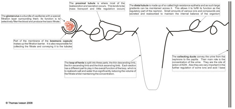
| + | |Body =<P ALIGN="left"> The nephron of the kidney is made up of two major parts; the renal corpuscle and the tubules. These are then both sub-divided into various parts and overall it is this structure which allows the kidney to filter the blood and then alter the composition of this filtrate to ensure that waste products are excreted and useful compounds preserved.</P> | ||
| + | <br> | ||
| + | <P ALIGN="left">The renal corpuscle can be subdivided into the glomerulus and the bowmans capsule. The tubules are split into the proximal tubule, the loop of henle, the distal tubule and the collecting ducts.</P> | ||
| + | |||
| + | <center>[[Image:nephovertri.jpg|800px]]</center> | ||
|subheading1colour = 66ff33 | |subheading1colour = 66ff33 | ||
| − | |subheading1 = [[ | + | |subheading1 = [[Nephron Microscopic Anatomy |Microscopic Anatomy of the Nephron]] |
|subheading1width =25 | |subheading1width =25 | ||
| − | |subheading1text = <center>[[ | + | |subheading1text = <center>[[Nephron Microscopic Anatomy #The Renal Corpuscle|The Renal Corpuscle]], [[Nephron Microscopic Anatomy #Proximal Tubule|Proximal Tubule]], [[Nephron Microscopic Anatomy #The Loop of Henle|The Loop of Henle]], [[Nephron Microscopic Anatomy #The Distal Tubule|The Distal Tubule]], [[Nephron Microscopic Anatomy #Collecting Duct|Collecting Duct]], [[Nephron Microscopic Anatomy #Blood Supply to the Nephron|Blood Supply to the Nephron]]</center> |
|subheading2colour = 66ff33 | |subheading2colour = 66ff33 | ||
| − | |subheading2 = [[ | + | |subheading2 = [[Urine Production (Table) - Anatomy & Physiology|Urine Production]] |
|subheading2width =25 | |subheading2width =25 | ||
|subheading2text = <center></center> | |subheading2text = <center></center> | ||
|subheading3colour = 66ff33 | |subheading3colour = 66ff33 | ||
| − | |subheading3 = [[ | + | |subheading3 = [[Water Balance and Homeostasis - Physiology|Water Balance and Homeostasis]] |
|subheading3width =25 | |subheading3width =25 | ||
|subheading3text = <center></Center> | |subheading3text = <center></Center> | ||
|subheading4colour = 66ff33 | |subheading4colour = 66ff33 | ||
| − | |subheading4 = [[ | + | |subheading4 = [[Essential Ion and Compound Balance and Homeostasis - Anatomy & Physiology|Essential Ion and Compound Balance and Homeostasis]] |
|subheading4width =25 | |subheading4width =25 | ||
|subheading4text = <center></Center> | |subheading4text = <center></Center> | ||
}} | }} | ||
| + | |||
| + | {{Learning | ||
| + | |flashcards = [[:Category:Urinary System Anatomy & Physiology Flashcards|Urinary System Anatomy Flashcards]] | ||
| + | }} | ||
| + | |||
| + | {{OpenPages}} | ||
| + | [[Category:Nephron]] | ||
Latest revision as of 14:16, 5 July 2012
| Nephron - Anatomy & Physiology Learning Resources | |
|---|---|
 Test your knowledge using flashcard type questions |
Urinary System Anatomy Flashcards |
Error in widget FBRecommend: unable to write file /var/www/wikivet.net/extensions/Widgets/compiled_templates/wrt6631abc350c471_35367738 Error in widget google+: unable to write file /var/www/wikivet.net/extensions/Widgets/compiled_templates/wrt6631abc3547536_29250856 Error in widget TwitterTweet: unable to write file /var/www/wikivet.net/extensions/Widgets/compiled_templates/wrt6631abc3576450_69345982
|
| WikiVet® Introduction - Help WikiVet - Report a Problem |
