Difference between revisions of "Serous Salivary Gland - Anatomy & Physiology"
Jump to navigation
Jump to search
| Line 1: | Line 1: | ||
| − | + | {{toplink | |
| − | + | |backcolour =BCED91 | |
| + | |linkpage =Alimentary - Anatomy & Physiology | ||
| + | |linktext =Alimentary System | ||
| + | |maplink = Alimentary (Concept Map)- Anatomy & Physiology | ||
| + | |pagetype =Anatomy | ||
| + | |sublink1=Oral Cavity - Salivary Glands - Anatomy & Physiology | ||
| + | |subtext1=SALIVARY GLANDS | ||
| + | }} | ||
| + | <br> | ||
'''Serous Salivary Glands''' | '''Serous Salivary Glands''' | ||
Revision as of 19:04, 20 September 2008
|
|
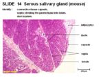
Serous Salivary Glands
- Connective tissue capsule
- Septa dividing the parenchyma into lobes
- Duct system
- Interlobular ducts run in the tissue septum lined by cuboidal to columnar epithelium
- Intralobular ducts run within the lobules. Striated intralobular ducts lined with cuboidal epithelium. Intercalated intralobular ducts lined with low cuboidal to simple squamous epithelium.
- Serous acini secrete a watery solution rich in proteins with spherical nuclei. Cells are pyramidal, cuboidal or crescent shaped.
