Difference between revisions of "Vascular Ring Anomalies"
| Line 1: | Line 1: | ||
{{review}} | {{review}} | ||
| − | + | ||
| − | |||
| − | |||
| − | |||
| − | |||
| − | |||
| − | |||
| − | |||
| − | |||
| − | |||
{{dog}}{{cat}} | {{dog}}{{cat}} | ||
[[Image:dextra-aorta.jpg|right|thumb|125px|<small><center>'''Dextra-aorta'''. Courtesy of A. Jefferies</center></small>]] | [[Image:dextra-aorta.jpg|right|thumb|125px|<small><center>'''Dextra-aorta'''. Courtesy of A. Jefferies</center></small>]] | ||
Revision as of 10:35, 22 June 2010
| This article has been peer reviewed but is awaiting expert review. If you would like to help with this, please see more information about expert reviewing. |

Most commonly found in dogs and cats. Acyanotic; clinical signs due to constriction of the oesophagus:
- Regurgiation of food, usually noticed at weaning.
- Aspiration pneumonia.
Persistent Right Aortic Arch
Most common vascular ring anomaly, increased incidence in GSDs, and Irish setters. vascular ring forms between the ductus arteriosus/ligamentum arteriosum and the persistent right aorta. Megaoesophagus is seen cranial to the constriction.
Other causes include:
Double aortic arch
Anomalous subclavian arteries
Arise form the aortic arch rather than the brachiocephalic artery.
Diagnosis:
- Ventral deviation of the trachea and dilation of the oesophagus on radiology.
- Barium swallow definitive.
Treatment:
- Surgical resection possible.
- Dietary management usually sufficient to control regurgitation.
- Seen most frequently in young animals right after weaning
- Common in dogs
- Very rare in cats
Signalment
- Genetics & Predisposed Breeds: Large breeds make up the majority of cases such as:
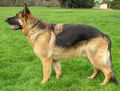 German Shepherd (Alsatian)
WikiCommons
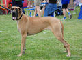 Great Dane
WikiCommons


Description
- During normal embryonic development there are five pairs of aortic arch arteries (1-6, 5 is absent) that undergo developmental changes necessary to form the major arteries of the head, neck, and upper thorax.
- Malformations of the aortic arch arteries can cause several vascular ring anomalies, but the most commonly seen anomaly is persistent right aortic arch (PRAA). With PRAA, the esophagus and trachea are most often encircled by the left ligamentum arteriosum as they pass over the heart base causing chronic esophageal and tracheal compression.
- When a young animal with PRAA is weaned onto solid food, the esophageal and tracheal compression causes food stasis above the narrowed esophageal foramen leading to megaesophagus. Continued stress on the esophagus in this manner can cause permanent damage. Additionally, aspiration pneumonia and the resulting respiratory distress is a complication of postprandial regurgitation.
Diagnosis
History & Clinical Signs
-Regurgitation (Postprandial=after meals)
-Coughing
-Respiratory distress
-Small size
-Underweight (Malnourished)
-Increased appetite
Physical Exam
-No cardiac murmurs unless PDA is also present
-Palpable dilated cervical esophagus
Radiographic Findings
-Megaesophagus in the thoracic inlet (+/- barium series)
Echocardiographic Findings
Not used to diagnose a PRAA
Electrocardiographic (ECG)
Not used to diagnose a PRAA
Treatment
-Surgical sectioning of the vascular ring anomaly to relieve constriction of the esophagus and trachea
-Management of esophageal damage: frequent feeding of small semi-solid/liquid meals from an elevated position
Prognosis
-Good with surgery
-Less favourable if there is extensive esophageal damage