Corpus Luteum Formation - Anatomy & Physiology
Introduction
Luteinisation occurs after ovulation and the collapse of the follicle. The number of corpora lutea formed in the ovary at any one time is directly proportional to the number of oocytes ovulated. Therefore many corpora lutea will be visible on the ovary of polytocous animals. During Luteinisation there is an increase in both the size and weight due to hyperplasia (increase in cell number) and hypertrophy (increase in cell size) within the developing corpus luteum.
Process of Luteinisation
Tissue Remodelling
- PGE2 released locally within the ovary causes activation of the body's tissue remodelling enzyme plasmin from its zymogen plasminogen. Activated Plasmin dissolves clot of the corpus Haemorrhagicum, formed due to disruption of follicular blood vessels at ovulation. Plasmin also helps in the remodelling of follicular basement membrane into copus luteum connective tissue.
- The organised follicle layers of Innner Granulosa cells and outer Theca cells is lost when the follicle implodes after ovulation. These cells become mixed within the copus luteum structure.
- There is an increase in the pigment lutein within the leteal cells.
- There is development of smooth endoplasmic reticulum within luteal cells due to the increase in steroid production.
Cell Differentiation
Two types of luteal cells are present within the corpus luteum:
Small luteal cells (<20µm)
- Formed from remodelled Follicular Theca cells. These cells proliferate during luteinisation.
- These cells contain many lipid droplets within their cytoplasm, an important source of cholesterol esters for Progesterone synthesis.
Large luteal cells (20-40µm)
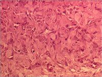
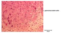
- Formed from Follicular Granulosa cells that have undergone hypertrophy.
- These large luteal cells are the endocrine cells of the corpus luteum producing large amounts of the hormone Progesterone.
- In some species secretory granules containing oxytocin or relaxin may be found close to the cell membrane.
Angiogenesis
- There is an increase cappilary cells and fibroblasts within the developing corpus luteum
- The extent to which the corpus luteum is vascularised may determine its functional capabilities as a large blood supply is required to provide adequate cholosterol for Progesterone synthesis.
Luteinisation Control
Control of corpus luteum formation and development as well as the production of Progesterone by luteal cells is regulated principally by Luteinising Hormone.
Error in widget FBRecommend: unable to write file /var/www/wikivet.net/extensions/Widgets/compiled_templates/wrt699faf7639cce0_39929412 Error in widget google+: unable to write file /var/www/wikivet.net/extensions/Widgets/compiled_templates/wrt699faf7647d025_66079690 Error in widget TwitterTweet: unable to write file /var/www/wikivet.net/extensions/Widgets/compiled_templates/wrt699faf764ed347_59447588
|
| WikiVet® Introduction - Help WikiVet - Report a Problem |

