Cytology Q&A 08
| This question was provided by Manson Publishing as part of the OVAL Project. See more Cytology Q&A. |
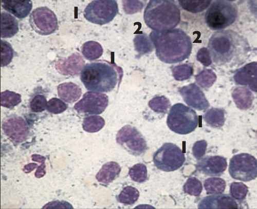
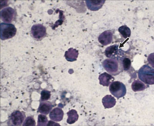
An eight-year-old crossbred dog has a history of crusting, flaky skin lesions, weight loss, lethargy and polyarthropathy. There is generalized, moderate lymphadenopathy. The dog was imported from Spain 18 months previously. Smears from FNAs from two lymph nodes are shown (both Giemsa, ×100 oil).
| Question | Answer | Article | |
| What cell(s) are increased in number? |
|
Link to Article | |
| What does this indicate? | This indicates reactive hyperplasia and particularly plasma cell hyperplasia. |
Link to Article | |
| What is the cell next to the arrowhead? | A ‘Mott cell’. This is a plasma cell containing Russell bodies, which are accumulations of immunoglobulins within vesicles. |
Link to Article | |
| Given the history and clinical signs, can you speculate on a possible cause for the generalized lymphadenopathy? | Plasma cell hyperplasia is caused by chronic antigenic stimulation. In this case the cause was Leishmania infection. The organisms were identified in macrophages in a bone marrow aspirate. Note:This case is included to emphasize recognition of basic processes and underlying mechanisms contributing to this cytological appearance. Knowledge of the significance of the finding of hyperplasia, with the history of importation from Spain, indicated that additional investigation was required to identify the suspected organism.
Therefore, an extensive work up may be required in some cases in order to determine the most likely underlying cause. The cytological evaluation is part of this work up. |
Link to Article | |

