Dental Analgesia and Anaesthesia
Introduction
The spectrum of patients requiring dental and oral surgery is wide and whilst most anaesthetic protocols may not vary much, special considerations should be made for this group of patients. Detection and treatment of pain can be challenging in this patient group as dental/oral examination may be limited in the conscious animal. It is only intra-operatively that a full assessment can be made and the degree of pathology, as well as treatment necessary and analgesia requirements can be fully established. Excellent pain management is essential to ensure good postoperative rehabilitation, especially to ensure optimal nutrition following dental or oral surgery.
Pain management should begin pre-operatively using an effective pre-emptive approach. This may however be difficult in some patients requiring oral and dental surgery, where significant pain can already exist. In this situation, the practice of multi-modal analgesia (MMA) is vital. Opioid analgesics form the basis of peri-operative MMA and are the most effective and commonly used analgesics for moderate to severe pain. Non-steroidal anti-inflammatory drugs (NSAIDs) are the most widely used analgesics in veterinary medicine and are also one of the best classes of drugs to treat and prevent post-operative pain. Care should be exercised in the timing of NSAID administration if blood loss and/or hypotension are anticipated.
Local anaesthesia prevents transmission of noxious stimuli, reducing the requirement for intra- and postoperative systemic analgesics. The use of loco-regional techniques is indicated as part of a balanced anaesthesia regimen for dental and oral surgery patients with the aim of providing pre-emptive analgesia. A great advantage is that by removing the noxious stimulation, less maintenance agent is needed to maintain an adequate level of anaesthesia, thus decreasing potential side effects. This has a particular advantage in maintaining adequate arterial blood pressure in for example, geriatric cats.
Specific Anaesthetic Considerations Regarding the Dental/Oral Patient
Geriatric Patients:
Owing to the relationship between age and the progression of periodontal and dental diseases, as well as the risk of oral neoplasia, geriatric patients often require dental and oral surgery procedures. There is often concurrent disease in these patients.
Young Patients:
Young patients may present with cleft palate and dental malocclusions. Considerations include fasting times, temperature management and management of blood loss.
Limited Oral Opening and Airway Access:
Temporo-mandibular joint disorders prevent oral opening to varying degrees, making orotracheal intubation difficult and sometimes even impossible. Other conditions such as masticatory myositis or craniomandibular osteopathy may also limit the extent to which the mouth can be opened, making access difficult.
Manifestation of Systemic Disease:
Dental disease may be a manifestation of systemic disease. Examples include patients with diabetes mellitus and patients with renal failure. Chronic dental disease may also compromise nutritional intake. An animal that is metabolically compromised is likely to have an increased anaesthetic risk.
Airway Management:
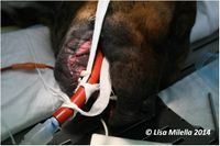
Cuffed endotracheal tubes (ETTs) will prevent gross debris from entering the airway, but will not prevent all liquid tracking down the trachea. Pack the oropharynx. The packing will become saturated and needs to be changed or squeezed out frequently to avoid oversaturation and water seepage into the airway. Lingual blood flow may become compromised if the pharynx is packed too tightly. Tie the throat pack to the ETT so that it is not accidentally left in situ at the end of the procedure. Care should be taken when inflating the ETT cuff, especially in cats, as over inflation is reported to cause tracheal rupture. Always turn the patient so that fluid does not run back into the mouth. The anaesthetic circuit should always be disconnected when turning to prevent movement of the ETT.
Hypothermia:
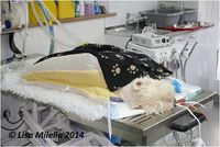
Heat loss starts at time of pre-anaesthetic medication, as the ability to compensate for heat loss is reduced. Temperature management is essential in all patients, but special attention should be paid to small, young, geriatric, thin or metabolically compromised animals. Heat loss is potentially high in dental and oral surgery patients as the mouth is always open and irrigation water used to cool instruments will also cool the patient. Care should be taken if the patient gets wet or hypotensive. Electric heat pads should be avoided if possible, and alternatives such as circulating warm water or hot air should be used.
Mouth Gags and Cats
Spring-held mouth gags can cause cerebral ischaemia and blindness in cats, and their use should be avoided. This risk may be compounded by hypotension and hypoxaemia. Gags can cause damage to the teeth, especially if the plane of anaesthesia is light and the animal begins to chew.
Loco-Regional Anaesthesia
The local anaesthetics most commonly used in veterinary dentistry belong to the aminoamide group. This includes lidocaine, mepivacaine, bupivacaine and ropivacaine. The choice of drug is usually dependent on the onset of action and duration of effect. The duration of effect may also depend on the site where the local anaesthesia is used – in very vascular areas, the drug may have less duration of action as it is resorbed quicker. The activity of local anaesthetics is dependent on the diffusion of the unionized form through the cell membrane, and the amount of unionized form is pH-dependent. Tissues with low pH will promote the presence of the ionized molecule, thus decreasing the action of the drug. Inflamed or infected tissues have a lower pH and thus the local anaesthetic agent may be less effective if injected into these tissues.
Bilateral or multiple blocks are often required, which impacts on the total volume available for use. For the selected drug, calculate the maximum safe dose and divide the volume available between the blocks planned.
The benefits of using local anaesthesia are well understood, however if poor technique is used these advantages are far outweighed by the potential risks and complications arising from its use. The greatest concern when injecting local anaesthetic is damage to a nerve or intravascular injection. Damage to the nerve can be minimized by using a fine (27gauge), short bevel needle with a gentle technique. The bevel should be orientated in the same direction as the nerve fibers in order to reduce the chance of cutting through the nerve fibers. When inserting the needle, avoid side to side movement, and if the needle touches bone, withdraw and replace the needle to avoid the damaged tip snagging on the nerve fibers. A smaller volume, injected slowly, should be used when injecting into a canal to avoid excessive pressure on the nerve to avoid neuropraxia. Penetration of a blood vessel is a frequent complication due to the presence of the large blood vessels in the neurovascular bundle. Using a fine needle in the way described above will help reduce the risk of a haematoma forming. Always aspirate before injecting the agent to reduce the risk of injecting intravascularly, which can lead to serious consequences.
The trigeminal nerve provides sensory innervations to the teeth and associated soft tissues. Knowledge of the applicable anatomy is useful to understand where to deposit the local anaesthetic agent to achieve the best results. Five of the most commonly used regional blocks are described below.
Mental Nerve Block
Superficial injection will anaesthetize the buccal soft tissues and lower lip rostral to the middle mental foramen. If the needle is introduced into the foramen, and advanced caudally, then sensation to the canine and incisors may be blocked. Care must be taken when inserting the needle so as not to cause damage to the neurovascular bundle. The preferred technique is to perform an inferior alveolar (mandibular) block to achieve anaesthesia for the mandibular teeth.
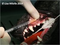
Technique: The middle mental foramen can be palpated through the lip frenulum just mesial to the 2nd premolar, in the ventral third of the mandible. The anaesthetic agent can be infused at the foramen and digital pressure applied to keep the agent local. The needle can also be introduced from a rostral to caudal direction, into the canal very carefully and the agent injected slowly. It is essential to aspirate prior to injecting. In small dogs and cats the canal is often too small to inject directly into it.
Inferior Alveolar Nerve Block
Anaesthetising the inferior alveolar nerve will provide loss of sensation of the hard and soft tissues of the ipsilateral mandible. The nerve enters the mandible at the level of the mandibular foramen. The mandibular nerve divides into three branches – the lingual nerve, the inferior alveolar nerve and the mylohyoid nerve. If the anaesthetic agent is deposited too far caudally, the other branches may also be affected, and may cause complications arising from loss of sensation to the tongue.
Technique: There are two approaches to this block – intra-oral or extra-oral. Either way, the mandibular foramen is palpated on the lingual aspect of the mandible, caudal and ventral to the last molar. It is not always possible to feel the foramen itself but usually the neurovascular bundle can be felt as a soft, string-like structure. When injecting intra-orally, the nerve or foramen is palpated with one hand whilst the syringe and needle are held in the other hand. The needle is advanced in a caudal direction, below the mucosa, with the bevel of the needle positioned towards the bone, to the position of the neurovascular bundle. Aspirate before injecting. For the extra-oral approach the neurovascular bundle is located as in the intra-oral approach and the needle is inserted perpendicularly, just medial to the body of the mandible, with the bevel facing the mandible. In the dog a useful landmark in helping to locate the position of where to insert the needle is the depression on the caudal border of the ventral mandible. The foramen can also be located halfway along an imaginary line drawn between the last molar and the angle of the jaw.
Infra-Orbital Nerve Block
This branch of the trigeminal nerve supplies the teeth and gingiva of the maxilla. The nerve exits from the infraorbital foramen which can be palpated just dorsal to the third premolar. An arterial pulse is often visible intra-orally at the foramen. Superficial injection at the foramen will anaesthetize the oral mucosa and upper lip rostral to the foramen. It will not anaesthetize the teeth. To anaesthetize the maxillary teeth, the needle should be inserted into the canal.
Technique: The needle is inserted into the foramen and advanced caudally with the needle parallel to the axis of the bone. In cats and brachycephalic dogs the canal may only be a few millimeters and inserting the needle into the canal may result in penetration of the eye. Always aspirate before injecting. There should be no resistance felt when injecting. Use a small volume, injected at low pressure to avoid neuropraxia.
Maxillary Nerve Block
This block anaesthetizes the infraorbital nerve as it enters the infraorbital canal. The block achieves anaesthesia of all of the maxillary teeth (including the last molar which is difficult to block with the intraorbital block), oral mucosa and buccal soft tissues and lip.
Technique: The block can be performed either intra-orally or extra-orally. For the intra-oral approach the needle is inserted behind the last molar, slightly palatal to the tooth. It is advanced at a slight angle rostrally. The extra-oral approach is the preferred approach – the landmark is the cranio-ventral border of the zygomatic arch. Feel the position of the last molar to gauge where to deposit the anaesthetic agent. The needle should be advanced from this point, parallel to the plane of the hard palate.
Palatine Nerve Block
The nerve is blocked at the level of the palatine foramen. It is difficult to palpate the foramen but the position is midway between the palatal root of the upper carnassial and the midline of the palate.
In summary, a basic dental analgesic plan:
- Include an opioid in the premedication.
- Use local anesthetics prior to surgery and/or administer additional opioids intraoperatively.
- Give opioids and/or NSAIDs postoperatively. Local anaesthesia (administered at the end of a procedure) will also provide postoperative analgesia.
- Administer NSAIDs during recovery.
| This article was written by Lisa Milella BVSc DipEVDC MRCVS. Date reviewed: 19 August 2014 |
| Endorsed by WALTHAM®, a leading authority in companion animal nutrition and wellbeing for over 50 years and the science institute for Mars Petcare. |
Error in widget FBRecommend: unable to write file /var/www/wikivet.net/extensions/Widgets/compiled_templates/wrt69a3d0aa4e3f06_77347135 Error in widget google+: unable to write file /var/www/wikivet.net/extensions/Widgets/compiled_templates/wrt69a3d0aa6523e8_50402943 Error in widget TwitterTweet: unable to write file /var/www/wikivet.net/extensions/Widgets/compiled_templates/wrt69a3d0aa7413c0_68603710
|
| WikiVet® Introduction - Help WikiVet - Report a Problem |


