Difference between revisions of "Reabsorption and Secretion Along the Proximal Tubule - Anatomy & Physiology"
m (→Sodium) |
|||
| (2 intermediate revisions by one other user not shown) | |||
| Line 1: | Line 1: | ||
| − | |||
==Introduction to Reabsorption== | ==Introduction to Reabsorption== | ||
| Line 65: | Line 64: | ||
===Sodium=== | ===Sodium=== | ||
| − | The majority (70%) of sodium is reabsorbed in the [[Reabsorption and Secretion Along the Proximal Tubule - Anatomy & Physiology| proximal tubule.]] It is reabsorbed into the cytosol of the epithelial cells either alone by [[Diffusion - Physiology| diffusion]] through [[Transport Proteins - Physiology#Diffusion Through Water Filled Protein Channels|ion channels]] followed by water and | + | The majority (70%) of sodium is reabsorbed in the [[Reabsorption and Secretion Along the Proximal Tubule - Anatomy & Physiology| proximal tubule.]] It is reabsorbed into the cytosol of the epithelial cells either alone by [[Diffusion - Physiology| diffusion]] through [[Transport Proteins - Physiology#Diffusion Through Water Filled Protein Channels|ion channels]] followed by water and chlorine or together with another product using a [[Transport Proteins - Physiology#Co-Transporters|co-transporter]]. |
| − | To maintain the concentration gradients and allow the diffusion to continue it is essential that sodium is not allowed to build up within the cell. This is the job of the sodium/potassium [[Transport Proteins - Physiology#ATPases|ATPase Pump]] and is an example of [[Active Transport - Physiology#Primary Active Transport|primary active transport]]. This pump removes | + | To maintain the concentration gradients and allow the diffusion to continue it is essential that sodium is not allowed to build up within the cell. This is the job of the sodium/potassium [[Transport Proteins - Physiology#ATPases|ATPase Pump]] and is an example of [[Active Transport - Physiology#Primary Active Transport|primary active transport]]. This pump removes sodium from the cell and puts potassium in. This creates a high concentration of potassium within the cell but this is corrected because there are also potassium ion channels in the basolateral membrane which allow potassium to diffuse back into the interstitium. Because both sodium and potassium are leaving the cell the net effect is that the tubular cells are negatively charged. This creates an electro gradient which further increases sodium uptake from the cells. The combined electrochemical gradient is very large allowing for great amounts of sodium to be reabsorbed. |
| − | Sodium is then able to move from the interstial fluid into the blood | + | Sodium is then able to move from the interstial fluid into the blood thanks to a combination of the blood having a low hydrostatic pressure and a high protein osmotic pressure. These conditions exist thanks to the [[Glomerular Apparatus and Filtration - Anatomy & Physiology#Factors Which Determine Selective Filtration|selective filtration]] of water, ions and glucose but the selective obstruction of proteins and promote the reabsorption of water and the associated dissolved ions within it back into the blood. |
===[[Aquaporins of the Kidney and Water Homeostasis - Anatomy & Physiology#The Ability of the Kidney To Alter the Water Content of the Body| Water]]=== | ===[[Aquaporins of the Kidney and Water Homeostasis - Anatomy & Physiology#The Ability of the Kidney To Alter the Water Content of the Body| Water]]=== | ||
| Line 143: | Line 142: | ||
Use the [[Reabsorption and Secretion Along the Proximal Tubule - Renal Flash Cards - Anatomy & Physiology|flash card revision resource]] for this section to test yourself. | Use the [[Reabsorption and Secretion Along the Proximal Tubule - Renal Flash Cards - Anatomy & Physiology|flash card revision resource]] for this section to test yourself. | ||
| − | + | ||
| − | [[Category: | + | [[Category:Nephron]] |
| − | |||
Revision as of 18:48, 9 December 2010
Introduction to Reabsorption
- The proximal tubule is a major site for reabsorption and some secretion.
- Gradients are small across the epithelium so tight regulation is not possible
- This occurs in the distal tubule
- 65-80% of the filtrate is reabsorbed
- Most reabsorption is coupled to sodium ion movement
- Small proteins and peptide hormones are reabsorbed by endocytosis
- Other substances are secreted or reabsorbed in the tubules passively by facilitated, non-facilitated diffusion or ion channels down chemical or electrical gradients.
- Other substances are secreted or reabsorbed by active transport against such gradients
- Movement is via ATPases.
- The reabsorbed material enters peritubular capillaries
- This is mainly driven by the Sodium/Pottasium ATPase in the basolateral membrane.
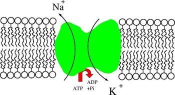
- This protein removes sodium from the cell maintaining the gradient between the lumen and the epithelium.
- Sodium reabsorption is driven by this protein
- Water and chloride then follow the transported sodium
- This is the most important transport mechanism in the proximal tubule
Proportion of Filtered Substances Reabsorbed in the Proximal Tubule
| Sodium and Water | |
| Organic solutes e.g. glucose and amino acids | |
| Potassium | |
| Urea | |
| Phosphate |
Epithelial Transport
Sodium is the most important ion in relation to reabsorption from the proximal tubule. Water and chloride follow sodium passively and many other ions, compounds and molecules are absorbed through co-transporters with sodium. However it is vital that intracellular levels of sodium remain low to favour this reabsorption so it falls to the sodium/potassium ATPase and sodium pump to remove sodium from the cell. Also to a lesser extent active transport of protons (H+). As well as directly sodium linked transport secondary active transport also plays a part however this does tend to be powered by sodium movement. Passive transport also has a role
Transport capacity is well above what is needed for normal plasma concentrations to ensure that adequate absorption occurs and that there is little/no wastage. As sodium, chloride and water are reabsorbed at the same rate, the filtrate concentrations remains the same along the proximal tubule. Only the volume of the filtrate decreases.
Ions and Compounds
The following ions and compounds are reabsorbed or secreted partly or completely in the proximal tubule:
Sodium
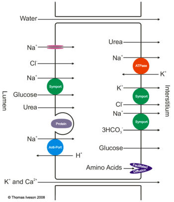
The majority (70%) of sodium is reabsorbed in the proximal tubule. It is reabsorbed into the cytosol of the epithelial cells either alone by diffusion through ion channels followed by water and chlorine or together with another product using a co-transporter.
To maintain the concentration gradients and allow the diffusion to continue it is essential that sodium is not allowed to build up within the cell. This is the job of the sodium/potassium ATPase Pump and is an example of primary active transport. This pump removes sodium from the cell and puts potassium in. This creates a high concentration of potassium within the cell but this is corrected because there are also potassium ion channels in the basolateral membrane which allow potassium to diffuse back into the interstitium. Because both sodium and potassium are leaving the cell the net effect is that the tubular cells are negatively charged. This creates an electro gradient which further increases sodium uptake from the cells. The combined electrochemical gradient is very large allowing for great amounts of sodium to be reabsorbed.
Sodium is then able to move from the interstial fluid into the blood thanks to a combination of the blood having a low hydrostatic pressure and a high protein osmotic pressure. These conditions exist thanks to the selective filtration of water, ions and glucose but the selective obstruction of proteins and promote the reabsorption of water and the associated dissolved ions within it back into the blood.
Water
Potassium
- Potassium is absorbed mainly by the paracellular route with water via osmosis
- Na+ / K+ ATPases in the basolateral membrane move potassium into epithelial cells from the interstitial spaces in order to remove sodium
- Potassium is then cleared from the cells using a co-transporter with chlorine
- No potassium excretion occurs here due to the lack of potassium ion channels in the apical membrane
Urea
50% of filtered urea is reabsorbed in the proximal tubule. However the concentration of urea actually increases thanks to the reabsorption of 70% of the filtered water in the same portion of the nephron. Urea is not able to be reabsorbed from this point until it reaches the lower portion of the collecting duct therefore its concentration further increases with the reabsorption of water.
Glucose
Glucose is a small molecule and so it is filtered in the same concentrations as are found in plasma which is approximately 5mmol/l. Reabsorption of glucose can only occur in the proximal tubule and occurs regardless of the concentration gradient as it is completed via secondary active transport. It is reabsorbed using a co-transporter with sodium. The realisation of the potential energy produced from sodium moving from an area of high concentration to an area of low concentration is enough energy to transport glucose across the membrane into the epithelial cells. The energy technically comes from the utilisation of ATP by the sodium/potassium ATPase which keeps sodium concentrations within the epithelial cells low this giving the sodium in the lumen a high potential energy.
Glucose is then passively transported out of the epithelial cells across the basolateral membrane.
It is normal that reabsorption is fully completed in the first half of this tubule however as the plasma concentration of glucose increases more of the tubule will be utilised to reabsorb the glucose. Concentrations of double the normal levels are required for glucose to appear in the urine and the concentration where glucose can first be detected is termed the renal threshold for glucose.
T Max and Splay
At approximately three times the normal levels the kidneys begin to approach the maximum possible reabsorption levels this is termed Tmax and is where the entire length of all the proximal tubules of all the nephrons of the kidney are working at maximum capacity.
Prior to this point the amount of glucose excreted does not increase linearly with the amount filtered. This is because some nephrons have longer proximal tubules than others so although some may be overcome and therefore allowing glucose to be excreted others are managing to fully reabsorb the glucose along their length. This results in what is termed splay. However after Tmax all the nephrons are at and above full capacity and therefore after this point any increase in filtered glucose is demonstrated linearly with that excreted.
The Tmax for a 20Kg dog is approximately 170mg per minute or 220g per 24 hour period. The normal amount of glucose filtered in 24 hours should be 60g.
The Kidneys Role In Glucose Regulation
The kidneys do not really regulate plasma glucose but their main aim is to preserve this valuable nutrient. It is only at very high levels which the kidneys play a role in helping to prevent any further rises in glucose via excretion.
The Secretion of H+ and the Reabsorption of HCO3-
Secretion of H+
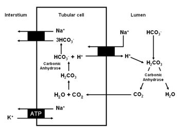
- The secretion of H+ in this section of the nephron is mainly a result of the Na+/H+ exchanger
- This is an antiporter in the apical membrane
- Energy for this process is provided by the Na/K ATPase in the basolateral membrane
- Therefore it is secondary active transport
- The ATPase pumps sodium out of the cell into the interstitium
- This maintains a low intracellular Na which creates a gradient for the absorption of sodium by the Na+/H+ antiporter
- This allows it to drive H against its concentration gradient
- Maintains a negative intracellular potential
- It is essential that HCO3- is removed from the cells by the co-transporter with sodium to ensure efficient H+ secretion.
Reabsorption of HCO3-
- Very efficient reabsorption mechanism
- 90% in first 1-2mm of tubule
- Lots of luminal carbonic anhydrase
- Stops the accumulation of H2CO3 in the lumen
- Keeps H+ concentration low - helps antiporter
- Roles of carbonic anhydrase
- In the cell forms HCO3- from OH and CO2
- In the tubule it works in reverse forming CO2 and H2O from the intermediate H2CO3 which forms from HCO3- and H+
- Allows continuous H+ secretion and HCO3- reabsorption
Protein
Peptide hormones and small amounts of albumin make it through the glomerular filtration barrier and these need to be reabsorbed. The reabsorption occurs via endocytosis in the proximal tubules. They are then broken down to amino acids in the epithelial cell cytoplasm and move via facilitated diffusion into the interstial fluid. The reabsorption of protein is usually complete though it is normal to detect small quantities of protein in the urine of some mammals e.g. the dog
Primary Active Secretion - Organic Acids and Bases
The secretion of these compounds occurs only in the proximal tubules. These molecules are mainly bound to plasma proteins with a small amount free in an active ionised form. It is only the free ions which are able to be transported. As the ionised molecules are transported out of the blood more molecules are ionised from the plasma proteins to take their place. These new ionised molecules are then able to be excreted thus releasing more and so it goes on. This allows a large amount of the substance to be secreted at one time. The mechanisms are not very selective and so many different substances are secreted at the same time. Secretion mechanisms are responsible for the secretion of drugs, hormones and things like food additives. Many unwanted or toxic organic molecules which enter the body are unionized. They therefore cannot be secreted so it falls to the liver to alter them into ionized forms to allow them to be disposed of.
Calcium
Half the plasma calcium is bound to proteins so it is only the ionised form which is available for filtration. Reabsorption of calcium occurs in the proximal tubule paracellulary however the regulation of how much is reabsorbed occurs in the ascending limb of the loop of henle, the distal tubule and collecting ducts.
Revision
Use the flash card revision resource for this section to test yourself.


