Difference between revisions of "Guttural Pouches - Anatomy & Physiology"
Fiorecastro (talk | contribs) |
|||
| (12 intermediate revisions by 4 users not shown) | |||
| Line 1: | Line 1: | ||
| − | |||
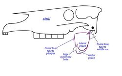
[[Image:Equine Guttural Pouch.jpg|thumb|right|250px|Equine Guttural Pouch - Copyright David Bainbridge]] | [[Image:Equine Guttural Pouch.jpg|thumb|right|250px|Equine Guttural Pouch - Copyright David Bainbridge]] | ||
Also known as: '''''Auditory Tube Diverticulum''''' | Also known as: '''''Auditory Tube Diverticulum''''' | ||
| Line 7: | Line 6: | ||
The Guttural Pouch is present only in members of the order Perissodactyla (nonruminant ungulates: horses, tapirs, rhinoceros) and another small band of small mammals including Hyraxes, certain bats and a South American mouse. | The Guttural Pouch is present only in members of the order Perissodactyla (nonruminant ungulates: horses, tapirs, rhinoceros) and another small band of small mammals including Hyraxes, certain bats and a South American mouse. | ||
| − | The guttural pouches are paired ventral diverticulae of the eustachian (auditory) tubes, formed by escape of mucosal lining of the tube through a relatively long ventral slit in the supporting cartilages. The auditory tube connect the [[Nasal Cavity - Anatomy & Physiology|nasal cavity]] and [[Ear - Anatomy & Physiology#Middle Ear|middle ear]] and the diverticulum dilates to form pouches which can have a capacity of 300-500ml in the domestic horse. The pouches are normally | + | The guttural pouches are paired ventral diverticulae of the eustachian (auditory) tubes, formed by escape of mucosal lining of the tube through a relatively long ventral slit in the supporting cartilages. The auditory tube connect the [[Nasal Cavity - Anatomy & Physiology|nasal cavity]] and [[Ear - Anatomy & Physiology#Middle Ear|middle ear]] and the diverticulum dilates to form pouches which can have a capacity of 300-500ml in the domestic horse. The pouches are normally aair filled. |
==Structure== | ==Structure== | ||
| Line 15: | Line 14: | ||
Right and left pouches are separated dorsomedially by rectus capitis ventralis and longus capitis muscles. Below this, by fused walls of the two pouches, the median septum is formed. | Right and left pouches are separated dorsomedially by rectus capitis ventralis and longus capitis muscles. Below this, by fused walls of the two pouches, the median septum is formed. | ||
| − | Each pouch is moulded to the stylohyoid | + | Each pouch is moulded to the stylohyoid muscle which divides the medial and lateral compartments, the medial compartment being approximately double the size of the lateral one and extends further caudally and ventrally. |
The guttural pouch has close association with many major structures including several [[Cranial Nerves - Anatomy & Physiology|cranial nerves]] (glossopharyngeal, vagus, accessory, hypoglossal), the sympathetic trunk and the external and internal carotid arteries. The pouch directly covers the temporohyoid joint. The pouch has an extremely thin wall which is lined by [[Respiratory Epithelium - Anatomy & Physiology|respiratory epithelium]] which secretes mucus. This normally drains into the pharynx when the horse is grazing. | The guttural pouch has close association with many major structures including several [[Cranial Nerves - Anatomy & Physiology|cranial nerves]] (glossopharyngeal, vagus, accessory, hypoglossal), the sympathetic trunk and the external and internal carotid arteries. The pouch directly covers the temporohyoid joint. The pouch has an extremely thin wall which is lined by [[Respiratory Epithelium - Anatomy & Physiology|respiratory epithelium]] which secretes mucus. This normally drains into the pharynx when the horse is grazing. | ||
| Line 35: | Line 34: | ||
Natural drainage of the pouch is throught the slit-like (pharyngeal) openings of the eustachian tube in the lateral wall of the nasopharynx. The connection opens when the horse swallows and grazing normally provides drainage. However, most of the pouch is ventral to his slit, and therefore drainage may be rather ineffective. If blocked, secretions accumulate and the pouch distends producing a palpable swelling. | Natural drainage of the pouch is throught the slit-like (pharyngeal) openings of the eustachian tube in the lateral wall of the nasopharynx. The connection opens when the horse swallows and grazing normally provides drainage. However, most of the pouch is ventral to his slit, and therefore drainage may be rather ineffective. If blocked, secretions accumulate and the pouch distends producing a palpable swelling. | ||
| − | == Function == | + | ==Function== |
| − | |||
| − | |||
| − | |||
| − | |||
| − | |||
| − | |||
| − | |||
| − | + | The Guttural Pouch is a mechanism for cooling the cerebral blood supply after exercise, due to its close connection with the internal carotid artery. | |
| − | |||
==References== | ==References== | ||
| Line 53: | Line 44: | ||
| − | [[Category:Respiratory System - Anatomy & Physiology | + | [[Category:Respiratory System - Anatomy & Physiology]] |
[[Category:A&P Done]] | [[Category:A&P Done]] | ||
Revision as of 19:23, 13 January 2011
Also known as: Auditory Tube Diverticulum
Introduction
The Guttural Pouch is present only in members of the order Perissodactyla (nonruminant ungulates: horses, tapirs, rhinoceros) and another small band of small mammals including Hyraxes, certain bats and a South American mouse.
The guttural pouches are paired ventral diverticulae of the eustachian (auditory) tubes, formed by escape of mucosal lining of the tube through a relatively long ventral slit in the supporting cartilages. The auditory tube connect the nasal cavity and middle ear and the diverticulum dilates to form pouches which can have a capacity of 300-500ml in the domestic horse. The pouches are normally aair filled.
Structure
The Guttural Pouch is located below the cranial cavity, towards the caudal end of the skull/wing of atlas. It is covered laterally by the Pterygoid muscles, parotid and mandibular glands. The floor lies mainly on the pharynx and beginning of the Oesophagus. The medial retropharyngeal lymph node lies between the pharynx and ventral wall of the pouches.
Right and left pouches are separated dorsomedially by rectus capitis ventralis and longus capitis muscles. Below this, by fused walls of the two pouches, the median septum is formed.
Each pouch is moulded to the stylohyoid muscle which divides the medial and lateral compartments, the medial compartment being approximately double the size of the lateral one and extends further caudally and ventrally.
The guttural pouch has close association with many major structures including several cranial nerves (glossopharyngeal, vagus, accessory, hypoglossal), the sympathetic trunk and the external and internal carotid arteries. The pouch directly covers the temporohyoid joint. The pouch has an extremely thin wall which is lined by respiratory epithelium which secretes mucus. This normally drains into the pharynx when the horse is grazing.
Several cranial nerves and arteries lie directly against the pouch as they pass to and from foramina in the caudal part of the skull (vessels within mucosal folds that indent the pouches):
Medial Compartment:
- Cranial nerves IX, X, XI, XII.
- Continuation of the sympathetic trunk beyond the cranial cervical ganglion.
- Internal carotid artery.
Lateral Compartment:
- Cranial nerve VII - limited contact with the dorsal part of the compartment.
- External carotid artery crosses the lateral wall of the lateral compartment in its approach (as maxillary artery) to the atlas canal. The external maxillary vein is also visible.
Drainage:
Natural drainage of the pouch is throught the slit-like (pharyngeal) openings of the eustachian tube in the lateral wall of the nasopharynx. The connection opens when the horse swallows and grazing normally provides drainage. However, most of the pouch is ventral to his slit, and therefore drainage may be rather ineffective. If blocked, secretions accumulate and the pouch distends producing a palpable swelling.
Function
The Guttural Pouch is a mechanism for cooling the cerebral blood supply after exercise, due to its close connection with the internal carotid artery.
References
Dyce, K.M., Sack, W.O. and Wensing, C.J.G. (2002) Textbook of Veterinary Anatomy. 3rd ed. Philadelphia: Saunders.
