Difference between revisions of "CNS Inflammation - Pathology"
Jump to navigation
Jump to search
(Redirected page to Category:Central Nervous System - Inflammatory Pathology) |
|||
| (4 intermediate revisions by the same user not shown) | |||
| Line 1: | Line 1: | ||
| − | # | + | [[Central Nervous System Inflammation Overview]] |
| + | |||
| + | ==Introduction== | ||
| + | |||
| + | * Although the CNS is well protected, its defences against organisms that have already invaded are less well developed. This is due to: | ||
| + | *# Minimal antibody production | ||
| + | *# Cerebrospinal fluid providing a good culture medium for invading organisms. | ||
| + | *# Inflammatory cell, antibody and drug entry to the CNS being impeded by the blood-brain barrier. | ||
| + | |||
| + | ===Classification of Inflammation=== | ||
| + | |||
| + | * CNS inflammation may manifest as encephalitis or meningitis. | ||
| + | ** These often co-exist. | ||
| + | * The aetiology CNS inflammation may be: | ||
| + | ** Infectious | ||
| + | *** Bacteria | ||
| + | *** Fungi | ||
| + | *** Protozoa | ||
| + | *** Viruses or non-infectious. | ||
| + | *** Infectious agents vary geographically. | ||
| + | ** Non-infectious | ||
| + | *** No infectious cause can be found in 60% of meningitis cases. | ||
| + | * Inflammation may also be broadly classified based on the nature of the exudate present. | ||
| + | ** '''Fibrinous''' | ||
| + | *** Caused by bacteria infection (including ''Mycoplasma''). | ||
| + | ** '''Suppurative''' | ||
| + | *** Caused by bacteria and fungi. | ||
| + | ** '''Granulomatous''' | ||
| + | *** Caused by bacteria or fungi. | ||
| + | ** '''Lymphoplasmacytic''' | ||
| + | *** Caused by viruses. | ||
| + | ** '''Haemorrhagic''' | ||
| + | *** This is rare. | ||
| + | *** Usually associated with septicemia or infarcts. | ||
| + | |||
| + | ==Clinical Signs of CNS Inflammation== | ||
| + | |||
| + | * Signs often reflect multiple levels of neurological involvement. | ||
| + | * Generalised [[Forebrain Disease - Pathology#Clinical Signs|forebrain signs]] are seen. | ||
| + | * Neck pain may be seen alone, or with other signs. | ||
| + | |||
| + | ==Diagnosis== | ||
| + | |||
| + | * History, physical and neurological examination. | ||
| + | * Fundic examination may give clues as to whether a systemic infection is present. | ||
| + | * CSF examination may help define the problem. | ||
| + | |||
| + | ==Treatment== | ||
| + | |||
| + | * Treatment is directed at a specific cause, if one can be found. | ||
| + | ** If a cause cannot be found, trimethoprim, clindamycin or doxycycline plus or minus corticosteroids may be used. | ||
| + | |||
| + | [[Category:Central Nervous System - Inflammatory Pathology]] | ||
| + | |||
| + | [[Central Nervous System Infectious Inflammation]] | ||
| + | |||
| + | ==Routes of Entry== | ||
| + | |||
| + | * CNS inflammation is usually the result of infection. | ||
| + | ** This may be caused by: | ||
| + | *** Bacteria | ||
| + | *** Fungi | ||
| + | *** Protozoa | ||
| + | *** Viruses | ||
| + | * Organisms must first enter the CNS in order to establish infection. | ||
| + | ** There are several routes of entry that allow this: | ||
| + | **# '''Haematogenous entry''' | ||
| + | **#* This is the most common route. | ||
| + | **# '''Entry via the peripheral nerves''' | ||
| + | **#* Organisms track within the axoplasm of axons. | ||
| + | **#* For example, ''Listeria monocytogenes''. | ||
| + | **# '''Penetrating trauma''' | ||
| + | **#* For example, dehorning wounds, skull fracture or tail docking. | ||
| + | **# '''Direct spread of infection''' | ||
| + | **#* From the nasal cavity, middle ear or paranasal sinuses. | ||
| + | |||
| + | ==Localisation of Infectious Organisms== | ||
| + | |||
| + | * After entry, organisms may establish in one or more of four main areas: | ||
| + | *# '''Epidural space''' | ||
| + | *#* Infection tends to manifest as abscess formation. | ||
| + | *# '''Subdural space''' | ||
| + | *#* Manifests as abscess formation. | ||
| + | *#* Fairly uncommon. | ||
| + | *# '''Leptomeninges''' | ||
| + | *#* Causes leptomeningitis, which may be: | ||
| + | *#*# Suppurative | ||
| + | *#*#* The most common form. | ||
| + | *#*#* Neutrophils are the predominant cell type. | ||
| + | *#*#* Caused by bacteria | ||
| + | *#*#** E.g. ''E. coli'' and ''Streptococcus'' | ||
| + | *#*#* There are often no gross lesions, but the brain may appear swollen and the meninges opaque. | ||
| + | *#*#* Usually results in death. | ||
| + | *#*# Eosinophilic meningoencephalitis | ||
| + | *#*#* The classic example of this is porcine salt poisoning, when water has been restricted and the suddenly replenished. | ||
| + | *#*#* Perivascular eosinophilic cuffing is seen in the cerebrum and meninges. | ||
| + | *#*# Lymphocytic | ||
| + | *#*#* Usually of viral origin. | ||
| + | *#*# Granulomatous | ||
| + | *#*#* Caused by fungal diseases and Mycobacteriosis. | ||
| + | *# '''CNS parenchyma''' | ||
| + | |||
| + | ==Bacterial Infections== | ||
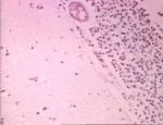
| + | [[Image:pneumococcalmeningitis.jpg|thumb|right|150px|Pneumococcal meningitis. Image courtesy of BioMed Archive]] | ||
| + | * Bacterial infections typically result in abscesses. | ||
| + | ** These may be single or multiple depending on the route of entry, and vary in size. | ||
| + | ** They contain a central, liquefied cavity. | ||
| + | * There are differences between cerebral abscesses and those occuring elsewhere. | ||
| + | ** Encapsulation is slow. | ||
| + | *** This is due to a lack of fibroblasts. | ||
| + | *** There is therefore less collagen in the capsule. | ||
| + | ** Astrocytic glial fibers are not as strong as collagen | ||
| + | * Other organisms may cause similar infections: | ||
| + | ** Rickettsial organisms | ||
| + | *** E.g. ''Ehrlichia'' | ||
| + | ** Spirochates | ||
| + | *** E.g. Leptospirosis | ||
| + | |||
| + | ==Viral Infections== | ||
| + | |||
| + | * Viral infections tend to reach the CNS by haematogenous spread and via peripheral nerves. | ||
| + | * There are three hallmark lesions of CNS viral infections: | ||
| + | *# Neuronal necrosis | ||
| + | *# Gliosis | ||
| + | *# Vascular changes | ||
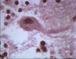
| + | * Several types of virus may cause inflammation in the CNS. [[Image:negribodies.jpg|thumb|right|150px|Negri bodies, as seen in rabies. Image courtesy of BioMed Archive]] | ||
| + | ** '''Neurotropic''', e.g. | ||
| + | *** Rabies (rhabdovirus) | ||
| + | *** Aujesky’s disease (herpesvirus) | ||
| + | *** Visna (ovine lentivirus) | ||
| + | ** '''Endotheliotropic''', e.g. | ||
| + | *** Infectious canine hepatitis (canine adenovirus) | ||
| + | *** Classical swine fever (pestivirus) | ||
| + | *** Equine herpesvirus type 1 (herpes) | ||
| + | ** '''Pantropic''' | ||
| + | *** Infectious canine distemper (morbillivirus) | ||
| + | *** Infectious bovine rhinotracheitis (bovine herpesvirus type 1) | ||
| + | * Other examples of viruses affecting the CNS: | ||
| + | ** Distemper | ||
| + | ** Parvovirus | ||
| + | ** Parainfluenza | ||
| + | ** Herpes | ||
| + | ** FIP | ||
| + | ** FIV | ||
| + | ** FeLV | ||
| + | ** Pseudorabies | ||
| + | ** Rabies | ||
| + | |||
| + | ==[[Prion Disease]]== | ||
| + | |||
| + | |||
| + | [[Category:Central Nervous System - Inflammatory Pathology]] | ||
| + | |||
| + | |||
| + | ==[[:Category:CNS Non-Infectious Inflammatory Diseases]]== | ||
| + | |||
| + | ===[[Granulomatous Meningoencephalitis]]=== | ||
| + | (GME) | ||
| + | * An [[CNS Idiopathic Conditions - Pathology|idiopathic CNS conditon]] | ||
| + | * May occur as: | ||
| + | ** A disseminated disease | ||
| + | ** A focal mass lesion | ||
| + | ** A primary occular disease | ||
| + | * Brainstem signs are common, although the forebrain is primarily affected. | ||
| + | * May be incorrectly diagnosed as lymphoma. | ||
| + | * Changes are apparent in the CSF. | ||
| + | ** There is usually a mononucloear pleocytosis. | ||
| + | ** Sometimes only protein is elveated. | ||
| + | * Diffuse inflammatory changes or a mass lesion will be seen by advanced imaging. | ||
| + | ** However, biopsy is required for a definative diagnosis. | ||
| + | * Life span is between 6 months and 1 year from diagnosis. | ||
| + | |||
| + | ====Treatment==== | ||
| + | |||
| + | * Immunosuppression: | ||
| + | ** Corticosteroids | ||
| + | ** Azathioprine | ||
| + | ** Cycophosphamide | ||
| + | * Surgery | ||
| + | ** This is only appropriate if there is a focal mass. | ||
| + | * Radiation therapy. | ||
| + | |||
| + | [[Necrotising Meningoencephalitis]] | ||
| + | |||
| + | [[:Category:CNS Non-Infectious Inflammatory Diseases]] | ||
| + | |||
| + | |||
| + | ===Pug Encephalitis=== | ||
| + | |||
| + | * A [[CNS Idiopathic Conditions - Pathology|CNS idiopathic condition]] | ||
| + | * Affects pugs. | ||
| + | ** Similar conditions are seen in yorkshire and maltese terriers. | ||
| + | * Officially known as necrotising meningoencephalitis of small dogs. | ||
| + | * Characterised by histological forebrain inflammation and necrosis. | ||
| + | * The disease is uniformly fatal. | ||
| + | ** Corticosterid treatment has no effect. | ||
| + | |||
| + | |||
| + | [:Category:CNS Non-Infectious Inflammatory Diseases]] | ||
Revision as of 12:51, 8 March 2011
Central Nervous System Inflammation Overview
Introduction
- Although the CNS is well protected, its defences against organisms that have already invaded are less well developed. This is due to:
- Minimal antibody production
- Cerebrospinal fluid providing a good culture medium for invading organisms.
- Inflammatory cell, antibody and drug entry to the CNS being impeded by the blood-brain barrier.
Classification of Inflammation
- CNS inflammation may manifest as encephalitis or meningitis.
- These often co-exist.
- The aetiology CNS inflammation may be:
- Infectious
- Bacteria
- Fungi
- Protozoa
- Viruses or non-infectious.
- Infectious agents vary geographically.
- Non-infectious
- No infectious cause can be found in 60% of meningitis cases.
- Infectious
- Inflammation may also be broadly classified based on the nature of the exudate present.
- Fibrinous
- Caused by bacteria infection (including Mycoplasma).
- Suppurative
- Caused by bacteria and fungi.
- Granulomatous
- Caused by bacteria or fungi.
- Lymphoplasmacytic
- Caused by viruses.
- Haemorrhagic
- This is rare.
- Usually associated with septicemia or infarcts.
- Fibrinous
Clinical Signs of CNS Inflammation
- Signs often reflect multiple levels of neurological involvement.
- Generalised forebrain signs are seen.
- Neck pain may be seen alone, or with other signs.
Diagnosis
- History, physical and neurological examination.
- Fundic examination may give clues as to whether a systemic infection is present.
- CSF examination may help define the problem.
Treatment
- Treatment is directed at a specific cause, if one can be found.
- If a cause cannot be found, trimethoprim, clindamycin or doxycycline plus or minus corticosteroids may be used.
Central Nervous System Infectious Inflammation
Routes of Entry
- CNS inflammation is usually the result of infection.
- This may be caused by:
- Bacteria
- Fungi
- Protozoa
- Viruses
- This may be caused by:
- Organisms must first enter the CNS in order to establish infection.
- There are several routes of entry that allow this:
- Haematogenous entry
- This is the most common route.
- Entry via the peripheral nerves
- Organisms track within the axoplasm of axons.
- For example, Listeria monocytogenes.
- Penetrating trauma
- For example, dehorning wounds, skull fracture or tail docking.
- Direct spread of infection
- From the nasal cavity, middle ear or paranasal sinuses.
- Haematogenous entry
- There are several routes of entry that allow this:
Localisation of Infectious Organisms
- After entry, organisms may establish in one or more of four main areas:
- Epidural space
- Infection tends to manifest as abscess formation.
- Subdural space
- Manifests as abscess formation.
- Fairly uncommon.
- Leptomeninges
- Causes leptomeningitis, which may be:
- Suppurative
- The most common form.
- Neutrophils are the predominant cell type.
- Caused by bacteria
- E.g. E. coli and Streptococcus
- There are often no gross lesions, but the brain may appear swollen and the meninges opaque.
- Usually results in death.
- Eosinophilic meningoencephalitis
- The classic example of this is porcine salt poisoning, when water has been restricted and the suddenly replenished.
- Perivascular eosinophilic cuffing is seen in the cerebrum and meninges.
- Lymphocytic
- Usually of viral origin.
- Granulomatous
- Caused by fungal diseases and Mycobacteriosis.
- Suppurative
- Causes leptomeningitis, which may be:
- CNS parenchyma
- Epidural space
Bacterial Infections
- Bacterial infections typically result in abscesses.
- These may be single or multiple depending on the route of entry, and vary in size.
- They contain a central, liquefied cavity.
- There are differences between cerebral abscesses and those occuring elsewhere.
- Encapsulation is slow.
- This is due to a lack of fibroblasts.
- There is therefore less collagen in the capsule.
- Astrocytic glial fibers are not as strong as collagen
- Encapsulation is slow.
- Other organisms may cause similar infections:
- Rickettsial organisms
- E.g. Ehrlichia
- Spirochates
- E.g. Leptospirosis
- Rickettsial organisms
Viral Infections
- Viral infections tend to reach the CNS by haematogenous spread and via peripheral nerves.
- There are three hallmark lesions of CNS viral infections:
- Neuronal necrosis
- Gliosis
- Vascular changes
- Several types of virus may cause inflammation in the CNS.
- Neurotropic, e.g.
- Rabies (rhabdovirus)
- Aujesky’s disease (herpesvirus)
- Visna (ovine lentivirus)
- Endotheliotropic, e.g.
- Infectious canine hepatitis (canine adenovirus)
- Classical swine fever (pestivirus)
- Equine herpesvirus type 1 (herpes)
- Pantropic
- Infectious canine distemper (morbillivirus)
- Infectious bovine rhinotracheitis (bovine herpesvirus type 1)
- Neurotropic, e.g.
- Other examples of viruses affecting the CNS:
- Distemper
- Parvovirus
- Parainfluenza
- Herpes
- FIP
- FIV
- FeLV
- Pseudorabies
- Rabies
Prion Disease
Category:CNS Non-Infectious Inflammatory Diseases
Granulomatous Meningoencephalitis
(GME)
- An idiopathic CNS conditon
- May occur as:
- A disseminated disease
- A focal mass lesion
- A primary occular disease
- Brainstem signs are common, although the forebrain is primarily affected.
- May be incorrectly diagnosed as lymphoma.
- Changes are apparent in the CSF.
- There is usually a mononucloear pleocytosis.
- Sometimes only protein is elveated.
- Diffuse inflammatory changes or a mass lesion will be seen by advanced imaging.
- However, biopsy is required for a definative diagnosis.
- Life span is between 6 months and 1 year from diagnosis.
Treatment
- Immunosuppression:
- Corticosteroids
- Azathioprine
- Cycophosphamide
- Surgery
- This is only appropriate if there is a focal mass.
- Radiation therapy.
Necrotising Meningoencephalitis
Category:CNS Non-Infectious Inflammatory Diseases
Pug Encephalitis
- A CNS idiopathic condition
- Affects pugs.
- Similar conditions are seen in yorkshire and maltese terriers.
- Officially known as necrotising meningoencephalitis of small dogs.
- Characterised by histological forebrain inflammation and necrosis.
- The disease is uniformly fatal.
- Corticosterid treatment has no effect.
[:Category:CNS Non-Infectious Inflammatory Diseases]]

