Difference between revisions of "Caecum - Anatomy & Physiology"
Fiorecastro (talk | contribs) |
(→Links) |
||
| (5 intermediate revisions by 5 users not shown) | |||
| Line 1: | Line 1: | ||
| − | |||
==Overview== | ==Overview== | ||
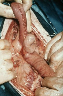
[[Image:opendogcaecum.jpg|thumb|right|250px|Dog caecum, ileum and colon - © RVC 2008]] | [[Image:opendogcaecum.jpg|thumb|right|250px|Dog caecum, ileum and colon - © RVC 2008]] | ||
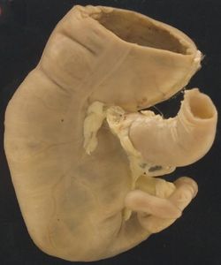
[[Image:Caecum and appendix of gibbon.JPG|thumb|right|250px|Gibbon caecum and appendix- © RVC 2008]] | [[Image:Caecum and appendix of gibbon.JPG|thumb|right|250px|Gibbon caecum and appendix- © RVC 2008]] | ||
| − | The | + | The caecum is a blind ending diverticulum of the large intestine and it exists at the junction of the '''[[Ileum - Anatomy & Physiology|ileum]]''' and the ascending '''[[Colon - Anatomy & Physiology|colon]]'''. Its size and physiological importance varies between species. It is a site of microbial fermentation, absorption and transportation. |
==Structure== | ==Structure== | ||
| Line 29: | Line 28: | ||
In the ruminant, the caecum is found on the right side of the abdomen, in the '''supraomental recess'''. The apex points caudally. It is relatively small and featureless, there are no taenia or haustra. Some microbial fermentation takes place. | In the ruminant, the caecum is found on the right side of the abdomen, in the '''supraomental recess'''. The apex points caudally. It is relatively small and featureless, there are no taenia or haustra. Some microbial fermentation takes place. | ||
| − | ===[[Alimentary System - | + | ===[[Equine Alimentary System - Anatomy & Physiology|Equine]]=== |
A significant amount of fermentation takes place in the equine caecum. | A significant amount of fermentation takes place in the equine caecum. | ||
| − | |||
===Porcine=== | ===Porcine=== | ||
| Line 48: | Line 46: | ||
{{Template:Learning | {{Template:Learning | ||
|flashcards = [[Caecum - Anatomy & Physiology - Flashcards|Caecum]] | |flashcards = [[Caecum - Anatomy & Physiology - Flashcards|Caecum]] | ||
| − | |videos = [ | + | |videos = [http://stream2.rvc.ac.uk/Anatomy/bovine/Pot0048.mp4 The Small and Large intestine of the Ruminant]<br>[http://stream2.rvc.ac.uk/Anatomy/bovine/Pot0052.mp4 Lateral view of the Abdomen of a young Ruminant]<br>[http://stream2.rvc.ac.uk/Anatomy/equine/Pony_abdomen.mp4 Lateral View of the Equine Abdomen]<br>[http://stream2.rvc.ac.uk/Frean/Pony/left_topography.mp4 Left Sided topography of the Equine abdomen]<br>[http://stream2.rvc.ac.uk/Frean/Pony/right_topography.mp4 Right sided topography of the Equine Abdomen]<br>[http://stream2.rvc.ac.uk/Anatomy/feline/pot0357.mp4 The Feline Abdomen]<br>[http://stream2.rvc.ac.uk/Frean/sheep/LargeSmallIntestine.mp4 Small and Large intestine of the Sheep]<br>[http://stream2.rvc.ac.uk/Anatomy/swine/Pig_abdomen.mp4 The Porcine Abdomen] |
}} | }} | ||
| − | |||
| − | |||
[[Category:Large Intestine - Anatomy & Physiology]] | [[Category:Large Intestine - Anatomy & Physiology]] | ||
[[Category:A&P Done]] | [[Category:A&P Done]] | ||
Revision as of 15:02, 23 May 2011
Overview
The caecum is a blind ending diverticulum of the large intestine and it exists at the junction of the ileum and the ascending colon. Its size and physiological importance varies between species. It is a site of microbial fermentation, absorption and transportation.
Structure
The caecum communicates with the ileum via the ileal orifice and with the colon via the caecocolic orifice. It consists of a base, body and apex. The apex is the blind-ending portion. It is attached to the ileum by a fold of peritoneum called the ileocaecal fold.
Function
1. Microbial Fermentation The contribution of this is species dependant. The products of fermentation are volatile fatty acids (VFAs).
2. Absorption VFAs that are produced are absorbed here.
3. Transportation Segmental contractions facilitate absorption and microbial activity. Every 3-5 minutes, segmentation is replaced by mass movements. This is similar to peristalsis, but large portions of the caecum contract simultaneously to move chyme into the colon.
Species Differences
Canine
In canine species, the caecum is on the right side of the abdomen. It is unique because it has no direct connection to the ileum. It is short and held in a spiral shape against the ileum by the ileocaecal fold. Little microbial fermentation takes place.
Ruminant
In the ruminant, the caecum is found on the right side of the abdomen, in the supraomental recess. The apex points caudally. It is relatively small and featureless, there are no taenia or haustra. Some microbial fermentation takes place.
Equine
A significant amount of fermentation takes place in the equine caecum.
Porcine
The caecum is on the left side of the abdomen, with the apex pointing caudoventrally. It is cylindrical in shape and there are three taenia present. The ventral taenia provides the attachment for the ileocaecal fold. The lateral and medial taenia are free.
Histology
The caecum has no villi. The mucosa has mucous glands, the lamina muscularis has large lymphatic nodules and the submucosa has no glands.
Links
Click here for information on the pathology of the Small and Large Intestines
Click here for information on the Peyer's Patches
| Caecum - Anatomy & Physiology Learning Resources | |
|---|---|
 Test your knowledge using flashcard type questions |
Caecum |
 Selection of relevant videos |
The Small and Large intestine of the Ruminant Lateral view of the Abdomen of a young Ruminant Lateral View of the Equine Abdomen Left Sided topography of the Equine abdomen Right sided topography of the Equine Abdomen The Feline Abdomen Small and Large intestine of the Sheep The Porcine Abdomen |

