Difference between revisions of "Reproductive Tract Prolapse"
| (One intermediate revision by one other user not shown) | |||
| Line 1: | Line 1: | ||
| − | |||
==Introduction== | ==Introduction== | ||
[[Image:Cervical Prolapse.jpg|thumb|right|200px|Prolapse,Copyright RVC 2008]] | [[Image:Cervical Prolapse.jpg|thumb|right|200px|Prolapse,Copyright RVC 2008]] | ||
| Line 39: | Line 38: | ||
==Uterine Prolapse== | ==Uterine Prolapse== | ||
This condition occurs '''after parturition''' and occurs sporadically in '''ewes, cattle, mares and sows'''. | This condition occurs '''after parturition''' and occurs sporadically in '''ewes, cattle, mares and sows'''. | ||
| − | |||
| − | |||
The condition may be complicated by '''prolapse of the bladder, uterine rupture or [[Hernia|intestinal herniation]]'''. One major concern is to prevent rupture of the '''large uterine vessels''' which will lead to rapid exsanguination. | The condition may be complicated by '''prolapse of the bladder, uterine rupture or [[Hernia|intestinal herniation]]'''. One major concern is to prevent rupture of the '''large uterine vessels''' which will lead to rapid exsanguination. | ||
| Line 49: | Line 46: | ||
===Clinical Signs=== | ===Clinical Signs=== | ||
The uterus protrudes out through the cervix, vagina and the lips of the vulva. The visible surface is the inside of the uterus and it may appear swollen, reddened and dry. There might also be tearing and bleeding and the placenta may still be partly attached to the uterine lining. | The uterus protrudes out through the cervix, vagina and the lips of the vulva. The visible surface is the inside of the uterus and it may appear swollen, reddened and dry. There might also be tearing and bleeding and the placenta may still be partly attached to the uterine lining. | ||
| − | |||
| − | |||
===Treatment=== | ===Treatment=== | ||
| − | |||
| − | |||
'''[[:Category:Fluid Therapy|Fluid therapy]]''' may be indicated if the animal is showing signs of shock and '''[[calcium]]''' might need to be administered. | '''[[:Category:Fluid Therapy|Fluid therapy]]''' may be indicated if the animal is showing signs of shock and '''[[calcium]]''' might need to be administered. | ||
| Line 82: | Line 75: | ||
A simple prolapse with retained placenta has a '''good prognosis'''. '''Subsequent breeding is not contraindicated''' and a prolapse is no more likely to occur again. The condition is not a reason to discontinue breeding the animal unless the uterus was damaged. | A simple prolapse with retained placenta has a '''good prognosis'''. '''Subsequent breeding is not contraindicated''' and a prolapse is no more likely to occur again. The condition is not a reason to discontinue breeding the animal unless the uterus was damaged. | ||
| − | |||
| − | |||
| − | |||
| − | |||
| − | |||
| − | |||
| − | |||
| − | |||
| − | |||
| − | |||
| − | |||
| − | |||
{{Learning | {{Learning | ||
| Line 99: | Line 80: | ||
[[Cattle Medicine Q&A 03]] | [[Cattle Medicine Q&A 03]] | ||
| − | |||
| − | |||
}} | }} | ||
| Line 111: | Line 90: | ||
Noakes, D. (2001) '''Arthur's veterinary reproduction and obstetrics''' ''Elsevier Health Sciences'' | Noakes, D. (2001) '''Arthur's veterinary reproduction and obstetrics''' ''Elsevier Health Sciences'' | ||
| − | |||
{{review}} | {{review}} | ||
| − | |||
| − | |||
[[Category:Expert Review]] | [[Category:Expert Review]] | ||
[[Category:Reproductive Disorders]][[Category:Parturition]] | [[Category:Reproductive Disorders]][[Category:Parturition]] | ||
[[Category:Reproductive Diseases - Horse]][[Category:Reproductive Diseases - Dog]] | [[Category:Reproductive Diseases - Horse]][[Category:Reproductive Diseases - Dog]] | ||
[[Category:Reproductive Diseases - Cattle]][[Category:Reproductive Diseases - Sheep]] | [[Category:Reproductive Diseases - Cattle]][[Category:Reproductive Diseases - Sheep]] | ||
Revision as of 15:09, 5 August 2011
Introduction
Prolapse of reproductive tract organs occurs in females during pregnancy or soon after parturition.
Cervical/vaginal prolapse is common in mature females in the last trimester, when the cervix is unable to support the foetus.
The vagina and/or cervix can also be expelled during delivery causing dystocia.
Uterine prolapse occurs early in the postpartum period and can contain the urinary bladder.
Vaginal Prolapse
Typically this is a disorder of ruminants occuring in late gestation. It occurs less frequently in pigs. In some bitches, hyperplasia of the vaginal mucosa occurs at oestrus, and sometimes protrudes through the vulva, but this is not comparable to the condition in the other species.
The condition is a welfare concern and can have a considerable economic impact in flocks that experience a high incidence.
Predisposing Factors
Hypocalcaemia, large foetal load, fat condition, inadequate exercise, short tail docking, excess dietary fibre, vaginal irritation, previous dystocia, inherited predisposition.
Clinical Signs
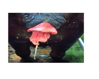
There is protrusion of varying parts of the vaginal wall and sometimes the cervix through the vulva so that the vaginal mucosa is exposed.
Animals might be clinically normal or show signs of exhaustion, shock and anaerobic infections.
Abortion or premature delivery, often of a dead foetus, may occur.
Acute intestinal herniation through the wall of the vagina may occur may be associated with vaginal prolapse and lead to death in affected animals.
Treatment
In all animals, caudal epidural anaesthesia will help improve the animal's pain and is an important welfare consideration. It will also facilitate replacement of the prolapse as the animal will stop straining.
The prolapse should be cleaned and the prolapse gently reduced by pushing with the palm of the hand. A very distended bladder should be allowed to empty with gentle pressure. In sheep, a vaginal retention device can be used such as a U-shaped device or a spoon attached to a harness or bailing twine.
Bühner's method of deep suturing of the vaginal wall can be used in sheep and cows. This returns the cervix to the correct position. The sutures can be loosened when signs of the prolapse resolve or parturition begins.
It is very important to replace the prolapse early to avoid any trauma and to ensure the animal maintains its pregnancy to term.
Uterine Prolapse
This condition occurs after parturition and occurs sporadically in ewes, cattle, mares and sows.
The condition may be complicated by prolapse of the bladder, uterine rupture or intestinal herniation. One major concern is to prevent rupture of the large uterine vessels which will lead to rapid exsanguination.
Predisposing Factors
Dystocia, forceful extraction of the foetus, retained placenta, vaginal lacerations and any condition causing straining after delivery.
Clinical Signs
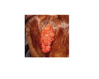
The uterus protrudes out through the cervix, vagina and the lips of the vulva. The visible surface is the inside of the uterus and it may appear swollen, reddened and dry. There might also be tearing and bleeding and the placenta may still be partly attached to the uterine lining.
Treatment
Fluid therapy may be indicated if the animal is showing signs of shock and calcium might need to be administered.
Epidural anaesthesia can be used to reduce straining and aid replacement of the prolapse.
Uterine tears may need to be sutured.
Ultrasonography can be used in mares to establish whether any intestine or the bladder is trapped within the prolapse. Urine may have to be aspirated from the bladder before it can be replaced.
The uterus should be thoroughly cleaned and copiously irrigated with saline. Lubricant can be applied to ease replacement.
Loose placenta can be removed but it should be left if it is firmly attached and if haemorrhage occurs.
To replace the prolapse, the animal should ideally be positioned with its hindquarters raised, in a frog-legged position. Initially the vaginal prolapse is replaced, followed by the cervix and finally the uterus. Cupped hands and gentle pressure should be used so that trauma is not caused to the friable tissue.
Once the uterus is replaced, the tips of the horns should be examined to assess if they have everted, and using a bottle to lengthen the arm, or filling the uterus with water can help.
After reduction is complete, low doses of oxytocin can be administered to facilitate rapid uterine involution.
Systemic antibiotics are essential due to the risk of metritis and non-steroidal antiinflammatory drugs serve to counteract endotoxaemia.
The placenta may be retained following uterine prolapse, and this should be treated subsequently.
Post-replacement, uterine involution should be assessed per rectum and antibiotics should be continued until placental membranes have passed and the uterus is involuting satisfactorily.
Death following prolapse is not uncommon, especially if haemorrhage occurs or help takes time to arrive.
A simple prolapse with retained placenta has a good prognosis. Subsequent breeding is not contraindicated and a prolapse is no more likely to occur again. The condition is not a reason to discontinue breeding the animal unless the uterus was damaged.
| Reproductive Tract Prolapse Learning Resources | |
|---|---|
 Test your knowledge using flashcard type questions |
Equine Reproduction and Stud Medicine Q&A 19 |
References
Aitken, I. (2007) Diseases of sheep Wiley-Blackwell
Knottenbelt, D. (2003) Equine stud farm medicine and surgery Elsevier Health Sciences
Robinson, N. (2009) Current therapy in equine medicine Elsevier Health Sciences
Noakes, D. (2001) Arthur's veterinary reproduction and obstetrics Elsevier Health Sciences
| This article has been peer reviewed but is awaiting expert review. If you would like to help with this, please see more information about expert reviewing. |

