Difference between revisions of "Innate Immunity Cellular Responses"
Jump to navigation
Jump to search
Rjfrancisrvc (talk | contribs) |
|||
| (23 intermediate revisions by 2 users not shown) | |||
| Line 1: | Line 1: | ||
==Introduction== | ==Introduction== | ||
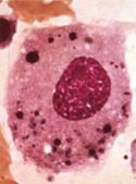
| − | [[Image:LH Macrophage Histology.jpg|thumb|right| | + | [[Image:LH Macrophage Histology.jpg|thumb|right|125px|<p>'''Macrophage'''</p><sup>© Nottingham Uni</sup>]] |
| − | Pathogens can invade the body if a breach occurs in the barriers formed by the skin and mucus membranes, for example a wound, they must be detected and destroyed by cellular and | + | Pathogens can invade the body if a breach occurs in the barriers formed by the skin and mucus membranes, for example a wound, they must be detected and destroyed by cellular and humoral means.<br /> |
<br /> | <br /> | ||
The cells involved in the cellular response to a wound are: | The cells involved in the cellular response to a wound are: | ||
| − | * Tissue | + | * Tissue mast cells and '''[[Macrtophages|macrophages]]''' that initially phagocytose and detect bacteria<br /> |
| + | <br /> | ||
| + | * The Blood granulocytes, or Polymorphonuclear (multi-lobed nuclei) Cells | ||
| + | ** The '''[[Neutrophils|Neutrophils]]''' are the most abundant as they are the primary cells that phagocytose bacteria, and the larger fungi | ||
| + | ** The '''[[Eosinophils|Eosinophils]]''' and '''[[Basophils|Basophils/ mast cells]]''' are only needed in rare circumstances as they are for killing parasites by the release of granules (exocytosis). | ||
<br /> | <br /> | ||
| − | |||
| − | |||
| − | |||
<br /> | <br /> | ||
* Blood '''[[Monocytes|monocytes]]''': phagocytose bacteria | * Blood '''[[Monocytes|monocytes]]''': phagocytose bacteria | ||
<br /> | <br /> | ||
| − | The main role of the innate cellular response is to delay systemic infection until the [[Adaptive Immune System|adaptive response]] can back it up with a more specific attack | + | <br /> |
| + | The main role of the innate cellular response is to delay systemic infection until the [[Adaptive Immune System|adaptive response]] can back it up with a more specific attack | ||
==[[Macrophages|Macrophages]]== | ==[[Macrophages|Macrophages]]== | ||
| − | [[Image:Monocytes.jpg|thumb|right| | + | [[Image:Monocytes.jpg|thumb|right|150px|Monocytes - J. Bredl, RVC 2008]] |
| − | The | + | *The role of macrophages in Innate Immunity is to act as primary '''phagocytes''' |
| − | * Alveolar macrophages | + | * Macrophages are present within tissues and take the form of distinct, tissue-specific populations: |
| − | * Tissue histiocytes | + | ** Alveolar macrophages |
| − | * Glomerular macrophages | + | ** Tissue histiocytes |
| − | * Hepatic Küpffer cells | + | ** Glomerular macrophages |
| − | * CNS microglia | + | ** Hepatic Küpffer cells |
| − | * Sinus-lining macrophages of the lymph nodes and spleen | + | ** CNS microglia |
| − | + | ** Sinus-lining macrophages of the lymph nodes and spleen | |
| − | + | * [[Monocytes|'''Monocytes''']] (immature macrophages) are circulating phagocytes | |
| − | + | ** Circulate for 6-8 hours | |
| − | + | ** Can function as phagocytes within the blood and as newly migrated cells in tissues | |
| − | + | ** Chiefly function to replace the various tissue macrophage populations | |
| − | |||
| − | |||
| − | |||
==[[Neutrophils|Neutrophils]]== | ==[[Neutrophils|Neutrophils]]== | ||
| − | [[Image:Neutrophil 2.jpg|thumb|right| | + | [[Image:Neutrophil 2.jpg|thumb|right|150px|Neutrophils - J. Bredl, RVC 2008]] |
| − | + | * Neutrophils are the principal, highly active '''phagocytes''' in the blood | |
| − | + | ** Comprise 30-70% of white blood cells depending on species | |
| − | + | ** Kill and digest microbes in a similar way as macrophages | |
| − | Neutrophils | + | * Neutrophils can also cause extracellular bacterial killing by disrupting bacterial membranes |
| − | + | ** Secrete small antibacterial peptides | |
| − | + | *** E.g. defensins and bactenecins | |
| − | + | * Neutrophils produce vasoactive peptides | |
| − | + | ** E.g. histamine and bradykinin | |
| − | + | ** Cause a great increase in extravasation of blood granulocytes and [[Monocytes|monocytes]] and plasma proteins at the site of infection | |
| − | + | * Neutrophils are the archetypal cell associated with [[:Category:Inflammation|acute inflammation]] | |
| − | + | ** Are attracted to sites of inflammation by: | |
| − | + | *** Complement activation | |
| − | Their removal from the site after the removal of infection is an important step in the resolution of the lesion | + | *** Cytokine production |
| + | *** Changes to vascular endothelium | ||
| + | ** Neutrophil activation in an inflammatory lesion results in the release of '''prostaglandins''' | ||
| + | *** Responsible for vasoactive changes and for pain | ||
| + | * The accumulation of dead and dying [[Neutrophils|neutrophils]] at the site of infection is called '''pus''' | ||
| + | ** Their removal from the site after the removal of infection is an important step in the resolution of the lesion | ||
==[[Eosinophils|Eosinophils]]== | ==[[Eosinophils|Eosinophils]]== | ||
| − | [[Image:Eosinophil.jpg|thumb|right| | + | [[Image:Eosinophil.jpg|thumb|right|150px|Eosinophil - J. Bredl, RVC 2008]] |
| − | Eosinophils are less common than [[Neutrophils|neutrophils]], | + | * Eosinophils are less common than [[Neutrophils|neutrophils]], and they are not phagocytic |
| − | + | ** Make up <5% of the leukocytes in normal blood | |
| − | + | * Eosinophil numbers are increased: | |
| − | + | ** Slightly during the resolution phase of inflammation | |
| − | + | ** Many-fold in parasite-infected animals | |
| − | + | *** The presence of a large proportion of eosinophils in a blood smear is highly indicative of parasitaemia | |
| − | + | * Mainly function by targeting the surface of parasites by means of specific antibody or complement | |
| − | + | ** Release a large range of toxic molecules that break down the parasite integument | |
| − | + | * Prominent in [[:Category:Allergic Diseases|allergic]] (anaphylactic) reactions | |
==[[Basophils|Basophils]] / [[Mast Cells|Mast Cells]]== | ==[[Basophils|Basophils]] / [[Mast Cells|Mast Cells]]== | ||
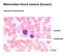
| − | [[Image:Basophil and Lymphocyte.jpg|thumb|right| | + | [[Image:Basophil and Lymphocyte.jpg|thumb|right|150px|Basophil - J. Bredl, RVC 2008]] |
| − | + | * Basophils/mast cells are principally localised at epithelial surfaces | |
| − | + | ** Very small numbers are present in blood | |
| − | + | *** Less than 0.5% circulating leukocytes | |
| − | + | * They have two principal functions: | |
| − | + | *# Induction of [[:Category:Inflammation|acute inflammation]] | |
| − | + | *#* Trauma and/ or bacterial infection causes the production of '''cytokines''' by the mast cells that induce a classical acute inflammatory response | |
| − | + | *# Response to parasite infection | |
| − | + | *#* Specific [[Immunoglobulins|IgE]] binds cells | |
| − | + | *#* Subsequent contact with antigen causes the mast cells to degranulate | |
| − | + | *#* Release enzymes and vasoactive substances that can result in a high level of mucus secretion and smooth muscle contraction | |
| − | + | * Also produce factors that influence local host cell physiology | |
| + | ** Various mediators increase the ratio of phagocyte to microbe | ||
<br><br> | <br><br> | ||
| − | |||
| − | |||
| − | |||
{{Jim Bee 2007}} | {{Jim Bee 2007}} | ||
[[Category:Innate Immune System]] | [[Category:Innate Immune System]] | ||
| − | |||
Revision as of 09:37, 1 May 2012
Introduction
Pathogens can invade the body if a breach occurs in the barriers formed by the skin and mucus membranes, for example a wound, they must be detected and destroyed by cellular and humoral means.
The cells involved in the cellular response to a wound are:
- Tissue mast cells and macrophages that initially phagocytose and detect bacteria
- The Blood granulocytes, or Polymorphonuclear (multi-lobed nuclei) Cells
- The Neutrophils are the most abundant as they are the primary cells that phagocytose bacteria, and the larger fungi
- The Eosinophils and Basophils/ mast cells are only needed in rare circumstances as they are for killing parasites by the release of granules (exocytosis).
- Blood monocytes: phagocytose bacteria
The main role of the innate cellular response is to delay systemic infection until the adaptive response can back it up with a more specific attack
Macrophages
- The role of macrophages in Innate Immunity is to act as primary phagocytes
- Macrophages are present within tissues and take the form of distinct, tissue-specific populations:
- Alveolar macrophages
- Tissue histiocytes
- Glomerular macrophages
- Hepatic Küpffer cells
- CNS microglia
- Sinus-lining macrophages of the lymph nodes and spleen
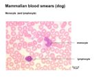
- Monocytes (immature macrophages) are circulating phagocytes
- Circulate for 6-8 hours
- Can function as phagocytes within the blood and as newly migrated cells in tissues
- Chiefly function to replace the various tissue macrophage populations
Neutrophils
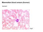
- Neutrophils are the principal, highly active phagocytes in the blood
- Comprise 30-70% of white blood cells depending on species
- Kill and digest microbes in a similar way as macrophages
- Neutrophils can also cause extracellular bacterial killing by disrupting bacterial membranes
- Secrete small antibacterial peptides
- E.g. defensins and bactenecins
- Secrete small antibacterial peptides
- Neutrophils produce vasoactive peptides
- E.g. histamine and bradykinin
- Cause a great increase in extravasation of blood granulocytes and monocytes and plasma proteins at the site of infection
- Neutrophils are the archetypal cell associated with acute inflammation
- Are attracted to sites of inflammation by:
- Complement activation
- Cytokine production
- Changes to vascular endothelium
- Neutrophil activation in an inflammatory lesion results in the release of prostaglandins
- Responsible for vasoactive changes and for pain
- Are attracted to sites of inflammation by:
- The accumulation of dead and dying neutrophils at the site of infection is called pus
- Their removal from the site after the removal of infection is an important step in the resolution of the lesion
Eosinophils
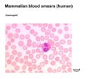
- Eosinophils are less common than neutrophils, and they are not phagocytic
- Make up <5% of the leukocytes in normal blood
- Eosinophil numbers are increased:
- Slightly during the resolution phase of inflammation
- Many-fold in parasite-infected animals
- The presence of a large proportion of eosinophils in a blood smear is highly indicative of parasitaemia
- Mainly function by targeting the surface of parasites by means of specific antibody or complement
- Release a large range of toxic molecules that break down the parasite integument
- Prominent in allergic (anaphylactic) reactions
Basophils / Mast Cells
- Basophils/mast cells are principally localised at epithelial surfaces
- Very small numbers are present in blood
- Less than 0.5% circulating leukocytes
- Very small numbers are present in blood
- They have two principal functions:
- Induction of acute inflammation
- Trauma and/ or bacterial infection causes the production of cytokines by the mast cells that induce a classical acute inflammatory response
- Response to parasite infection
- Specific IgE binds cells
- Subsequent contact with antigen causes the mast cells to degranulate
- Release enzymes and vasoactive substances that can result in a high level of mucus secretion and smooth muscle contraction
- Induction of acute inflammation
- Also produce factors that influence local host cell physiology
- Various mediators increase the ratio of phagocyte to microbe
| Originally funded by the RVC Jim Bee Award 2007 |
