Difference between revisions of "Spermatogenesis and Spermiation - Anatomy & Physiology"
Fiorecastro (talk | contribs) |
RebekahMCB (talk | contribs) |
||
| (12 intermediate revisions by 3 users not shown) | |||
| Line 1: | Line 1: | ||
| − | {{ | + | {{review}} |
| + | |||
==Introduction== | ==Introduction== | ||
| − | + | Spermatogenesis is the process of the gradual transformation of germ cells into spermatozoa. It occurs mainly within the seminiferous tubules of the testes and can be divided into three phases, each of which is associated with different germ cell types: | |
| − | |||
| − | Spermatogenesis is the process of the gradual transformation of germ cells into spermatozoa. It occurs mainly within the | ||
| − | |||
| − | |||
| − | |||
| − | |||
| − | |||
| − | + | *Proliferative phase - spermatogonia → spermatocytes | |
| + | *Meiotic phase - spermatocytes → spermatids | ||
| + | *Differentiation phase / spermiogenesis - spermatids → spermatozoa | ||
| − | + | Unlike the female production of gametes which occurs entirely before birth, with gamete maturation occurring in a pulsatile fashion after puberty, males produce gametes continuously from puberty onwards for the rest of their reproductive lives and the release of the gametes is constant. | |
| − | |||
| − | + | ==The seminiferous tubules== | |
| + | [[Image:spermatogenesis.jpg|thumb|right|150px|Spermatogenesis Copyright Amy Cartmel 2008]] | ||
| + | The seminiferous tubules are the site of spermatogenesis. The 2 main cell types within the tubules involved in spermatogenesis are the germ cells, which will develop into sperm, and somatic cells known as Sertoli cells, which nuture the germ cells throughout the development process. | ||
| − | + | As the germ cells progress through their stages of development they move slowly from the basement membrane of the tubules through the tight junction of the Sertoli cells into the tubular lumen. | |
| − | |||
| − | |||
| − | + | =Stages of spermatogenesis= | |
| + | [[File:Spermatogenesis diagram v2.jpg|thumb|Sperm developing within seminiferous tubule]] | ||
| + | ==The proliferation phase== | ||
| + | Stem or A '''spermatogonia''' located in the basal region of the tubular epithelium undergo mitosis. The progeny of these divisions maintain their own numbers as well as giving rise to several interconnected B spermatogonia (the number of these arising from a single A spermatogonia is species dependent). B spermatogonia divide to give rise to primary (1<sup>o</sup>) '''spermatocytes'''. All descendants of a B spermatogonium remain connected by cytoplasmic bridges, forming a syncytium - like cell clone which undergoes synchronous development. | ||
| − | == | + | ==The meiotic phase== |
| − | Each 1<sup>o</sup> spermatocyte divides to give rise to two short-lived | + | Each 1<sup>o</sup> spermatocyte divides to give rise to two short-lived 2<sup>o</sup> spermatocytes, which in turn give rise to two '''spermatids''' each, each of which contain a haploid number of chromosomes (half the number of a somatic cell). |
1<sup>o</sup> spermatocytes are the largest cells in the spermatogenic series and are located approximately midway within the seminiferous epithelium. | 1<sup>o</sup> spermatocytes are the largest cells in the spermatogenic series and are located approximately midway within the seminiferous epithelium. | ||
| − | The process of meiosis occurs over a long period, with prophase of the first meiotic division taking up to | + | The process of meiosis occurs over a long period, with prophase of the first meiotic division taking up to 3 weeks <ref name="Hess">Hess RA, Spermatogenesis, overview. In: Knobil E, Neil JD (eds.), Encyclopedia of Reproduction. New York: Academic Press; 1999: pp. 539–545</ref> |
| − | == | + | ==The differentiation phase== |
| − | This phase is also known as | + | This phase is also known as spermiogenesis. |
| − | Spermatids undergo transformation into | + | Spermatids undergo transformation into spermatozoa. Many changes occur within the cells, the 3 major ones being |
| − | + | i) formation of the acrosome, which covers the cranial part of the head. The acrosome will contain hydrolytic enzymes to allow fusion of sperm and egg for fertilisation. | |
| − | i) formation of the | ||
| − | ii) | + | ii) condensation of nuclear chromatin in the head to form a dark-staining structure |
| − | iii) | + | iii) growth of the tail opposite the acrosome, and loss of excess cytoplasmic material which is shed as a residual body. The body is phagoctosed by the Sertoli cells. |
The morphological changes occurring during this process can be seen if sections of different seminiferous tubules are examined. | The morphological changes occurring during this process can be seen if sections of different seminiferous tubules are examined. | ||
| − | + | =Hormonal Control of spermatogenesis= | |
| − | + | The production of sperm is controlled by hormones influencing sertoli cells rather than sperm cells directly. Hormonal control is via the two gonadotrophins luteinising hormone (LH) and follicle-stimulating hormone (FSH). | |
| − | + | *LH acts on the interstitial leydig cells stimulating them to produce the androgen testosterone. | |
| − | + | *FSH acts on the sertoli cells within the seminipherous tubules stimulating production of Androgen Binding Protein as well as Inhibin | |
| − | + | <br> | |
| − | |||
| − | |||
| − | |||
| − | |||
| − | |||
| − | |||
| − | FSH acts on the | ||
| − | |||
| − | Inhibin | ||
| − | |||
| − | |||
| − | |||
| − | |||
| − | |||
| − | |||
| − | |||
| − | |||
| − | |||
| − | |||
<references/> | <references/> | ||
| − | |||
| − | |||
| − | |||
| − | |||
{{Template:Learning | {{Template:Learning | ||
| − | |videos = [[ | + | |videos = [[Equine Testes video|Dissection of the equine testicle]] |
|powerpoints = [[Male Reproductive Tract Histology resource|Histology of the Male reproductive tract]] | |powerpoints = [[Male Reproductive Tract Histology resource|Histology of the Male reproductive tract]] | ||
}} | }} | ||
| − | |||
| − | |||
| − | |||
[[Category:Male Reproduction]] | [[Category:Male Reproduction]] | ||
Revision as of 20:40, 4 May 2012
| This article has been peer reviewed but is awaiting expert review. If you would like to help with this, please see more information about expert reviewing. |
Introduction
Spermatogenesis is the process of the gradual transformation of germ cells into spermatozoa. It occurs mainly within the seminiferous tubules of the testes and can be divided into three phases, each of which is associated with different germ cell types:
- Proliferative phase - spermatogonia → spermatocytes
- Meiotic phase - spermatocytes → spermatids
- Differentiation phase / spermiogenesis - spermatids → spermatozoa
Unlike the female production of gametes which occurs entirely before birth, with gamete maturation occurring in a pulsatile fashion after puberty, males produce gametes continuously from puberty onwards for the rest of their reproductive lives and the release of the gametes is constant.
The seminiferous tubules
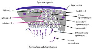
The seminiferous tubules are the site of spermatogenesis. The 2 main cell types within the tubules involved in spermatogenesis are the germ cells, which will develop into sperm, and somatic cells known as Sertoli cells, which nuture the germ cells throughout the development process.
As the germ cells progress through their stages of development they move slowly from the basement membrane of the tubules through the tight junction of the Sertoli cells into the tubular lumen.
Stages of spermatogenesis
The proliferation phase
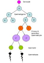
Stem or A spermatogonia located in the basal region of the tubular epithelium undergo mitosis. The progeny of these divisions maintain their own numbers as well as giving rise to several interconnected B spermatogonia (the number of these arising from a single A spermatogonia is species dependent). B spermatogonia divide to give rise to primary (1o) spermatocytes. All descendants of a B spermatogonium remain connected by cytoplasmic bridges, forming a syncytium - like cell clone which undergoes synchronous development.
The meiotic phase
Each 1o spermatocyte divides to give rise to two short-lived 2o spermatocytes, which in turn give rise to two spermatids each, each of which contain a haploid number of chromosomes (half the number of a somatic cell). 1o spermatocytes are the largest cells in the spermatogenic series and are located approximately midway within the seminiferous epithelium.
The process of meiosis occurs over a long period, with prophase of the first meiotic division taking up to 3 weeks [1]
The differentiation phase
This phase is also known as spermiogenesis.
Spermatids undergo transformation into spermatozoa. Many changes occur within the cells, the 3 major ones being i) formation of the acrosome, which covers the cranial part of the head. The acrosome will contain hydrolytic enzymes to allow fusion of sperm and egg for fertilisation.
ii) condensation of nuclear chromatin in the head to form a dark-staining structure
iii) growth of the tail opposite the acrosome, and loss of excess cytoplasmic material which is shed as a residual body. The body is phagoctosed by the Sertoli cells.
The morphological changes occurring during this process can be seen if sections of different seminiferous tubules are examined.
Hormonal Control of spermatogenesis
The production of sperm is controlled by hormones influencing sertoli cells rather than sperm cells directly. Hormonal control is via the two gonadotrophins luteinising hormone (LH) and follicle-stimulating hormone (FSH).
- LH acts on the interstitial leydig cells stimulating them to produce the androgen testosterone.
- FSH acts on the sertoli cells within the seminipherous tubules stimulating production of Androgen Binding Protein as well as Inhibin
- ↑ Hess RA, Spermatogenesis, overview. In: Knobil E, Neil JD (eds.), Encyclopedia of Reproduction. New York: Academic Press; 1999: pp. 539–545
| Spermatogenesis and Spermiation - Anatomy & Physiology Learning Resources | |
|---|---|
 Selection of relevant videos |
Dissection of the equine testicle |
 Selection of relevant PowerPoint tutorials |
Histology of the Male reproductive tract |

