Difference between revisions of "Mast Cells"
Rjfrancisrvc (talk | contribs) |
|||
| (9 intermediate revisions by 4 users not shown) | |||
| Line 2: | Line 2: | ||
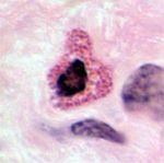
[[Image:LH Mast cells Histology.jpg|right|thumb|150px|<p>'''Mast cells'''</p><sup>©Nottingham Uni 2008</sup>]] | [[Image:LH Mast cells Histology.jpg|right|thumb|150px|<p>'''Mast cells'''</p><sup>©Nottingham Uni 2008</sup>]] | ||
==Introduction== | ==Introduction== | ||
| − | <p>Mast cells are derived from the same stem cell line as [[Basophils|basophils]] and while they are similar to | + | <p>Mast cells are derived from the same stem cell line as [[Basophils|basophils]] and while they are similar to basophils they are not identical. Mast cells are found in connective tissue and are larger than other [[Leukocytes|leukocytes]] at 15-20µm. They have cytoplasm containing a high number of large, intensely staining basophilic granules which mask the nucleus.</p> |
==Development== | ==Development== | ||
| − | <p>Mast cells CFU | + | <p>Mast cells colony forming units (CFU) develop in the [[Bone Marrow - Anatomy & Physiology|bone marrow]] from the myeloid stem cell ([[Haematopoiesis - Overview#Colony Forming Units|CFU-GEMM]]) under the stimulation of IL-3 and Stem Cell Factor (SCF). Then under the continual stimulation with SCF, and with the additional stimulation with IL-6, the immature mast cells are formed. These then enter circulation where they remain undifferentiated. On migration into tissue they then differentiate, under stimulation by IL-4, and produce their granules. Once in the tissue mast cells may last for weeks to years.</p> |
==Granules== | ==Granules== | ||
| Line 10: | Line 10: | ||
* Preformed mediators such as serotonin, histamine, proteoglycans, heparin and serine proteases (tryptase and/or chymase) | * Preformed mediators such as serotonin, histamine, proteoglycans, heparin and serine proteases (tryptase and/or chymase) | ||
* Lipid mediators such as leukotrienes and prostaglandin D2 | * Lipid mediators such as leukotrienes and prostaglandin D2 | ||
| − | * Cytokines: | + | * Cytokines: IL-3, IL-4, IL-5, IL-6, IL-8, IL-13, IL-16 and TNF-α</p> |
| − | *Chemotactic factors for both [[Neutrophils|neutrophils]] and [[Eosinophils|eosinophils]] | + | * Chemotactic factors for both [[Neutrophils|neutrophils]] and [[Eosinophils|eosinophils]] |
<p>Histamine increases vascular permeability, mucous production and smooth muscle tone in the respiratory tract and is responsible for the itch sensation in the skin. Heparin is an anticoagulant and binds with antithrombin III and platelet factor IV to block coagulation factors. Leukotrienes like histamine are responsible for contraction of respiratory smooth muscle. Chymase plays a role in the production of angiotensin II.</p> | <p>Histamine increases vascular permeability, mucous production and smooth muscle tone in the respiratory tract and is responsible for the itch sensation in the skin. Heparin is an anticoagulant and binds with antithrombin III and platelet factor IV to block coagulation factors. Leukotrienes like histamine are responsible for contraction of respiratory smooth muscle. Chymase plays a role in the production of angiotensin II.</p> | ||
| − | <p> | + | <p>Mast Cell granules are thought to produce a ground substance to facilitate collagen formation which occurs during the repair of an injury.</p> |
==Actions== | ==Actions== | ||
| Line 21: | Line 21: | ||
==In pathology== | ==In pathology== | ||
*Classically a cell involved in [[Acute Inflammation|acute inflammation]] | *Classically a cell involved in [[Acute Inflammation|acute inflammation]] | ||
| − | *Mast cell tumours of the intestine and [[Mast | + | *[[Neoplasia_-_Pathology#Mast_Cell_Proliferation_Disorders|Mast cell tumours]] of the intestine, liver and skin. |
| + | |||
| + | {{Template:Learning | ||
| + | |powerpoints = [[Connective Tissue Histology resource|Tutorial about connective tissues, with slides on Mast Cells]] | ||
| + | }} | ||
| + | |||
==References== | ==References== | ||
<references /> | <references /> | ||
<br> | <br> | ||
| − | [[Category:Blood_Cells]] | + | [[Category:Blood_Cells]] [[Category:Kate English reviewing]] |
Latest revision as of 14:32, 22 May 2012
Introduction
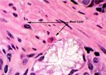
Mast cells are derived from the same stem cell line as basophils and while they are similar to basophils they are not identical. Mast cells are found in connective tissue and are larger than other leukocytes at 15-20µm. They have cytoplasm containing a high number of large, intensely staining basophilic granules which mask the nucleus.
Development
Mast cells colony forming units (CFU) develop in the bone marrow from the myeloid stem cell (CFU-GEMM) under the stimulation of IL-3 and Stem Cell Factor (SCF). Then under the continual stimulation with SCF, and with the additional stimulation with IL-6, the immature mast cells are formed. These then enter circulation where they remain undifferentiated. On migration into tissue they then differentiate, under stimulation by IL-4, and produce their granules. Once in the tissue mast cells may last for weeks to years.
Granules
Mast cells can be classified by the presence or lack of tryptase and/or chymase. MCTC mast cells have granules that contain tryptase and chymase (found mainly in skin and intestinal submucosa) while MCT mast cells have granules containing only tryptase (found mainly in respiratory tissue and intestinal mucosa). They can also be classified by their location within the body[1]. Mast cells in connective tissue are larger, have the proteoglycan heparin and contain high amounts of histamine while mucosal mast cells contain less histamine and have the proteoglycan chondroitin sulphate.
More generally the granules contain:
- Preformed mediators such as serotonin, histamine, proteoglycans, heparin and serine proteases (tryptase and/or chymase)
- Lipid mediators such as leukotrienes and prostaglandin D2
- Cytokines: IL-3, IL-4, IL-5, IL-6, IL-8, IL-13, IL-16 and TNF-α
- Chemotactic factors for both neutrophils and eosinophils
Histamine increases vascular permeability, mucous production and smooth muscle tone in the respiratory tract and is responsible for the itch sensation in the skin. Heparin is an anticoagulant and binds with antithrombin III and platelet factor IV to block coagulation factors. Leukotrienes like histamine are responsible for contraction of respiratory smooth muscle. Chymase plays a role in the production of angiotensin II.
Mast Cell granules are thought to produce a ground substance to facilitate collagen formation which occurs during the repair of an injury.
Actions
Mast cells are involved in the inflammatory response and play a key part in anaphylaxis.
IgE released by plasma cells in response to antigens bind to FC receptors on the mast cell surface. This stimulates the mast cells to degranulate. The granule contents cause blood vessel permeability to increase and, along with chemotactic factors, this enhances the migration of other leukocytes to the area. The production of angiotensin II by chymase causes systemic vasoconstriction in response to vascular damage.
In pathology
- Classically a cell involved in acute inflammation
- Mast cell tumours of the intestine, liver and skin.
| Mast Cells Learning Resources | |
|---|---|
 Selection of relevant PowerPoint tutorials |
Tutorial about connective tissues, with slides on Mast Cells |
References
- ↑ Metcalfe, D.D., Baram, D. and Mekori, Y. (1997) Mast Cells. Physiological Reviews 77(4): pp.1033-1064.

