Difference between revisions of "Vascular Ring Anomalies"
m (Text replace - "Category:Cardiovascular Pathology - Dog" to "Category:Cardiovascular Diseases - Dog") |
TestStudent (talk | contribs) |
||
| (17 intermediate revisions by 3 users not shown) | |||
| Line 1: | Line 1: | ||
| − | + | {{OpenPagesTop}} | |
| − | + | Conditions included: '''''Persistent Right Aortic Arch (PRAA) — Double Aortic Arch — Anomalous Subclavian Arteries''''' | |
| − | |||
| − | ==== | + | == Introduction== |
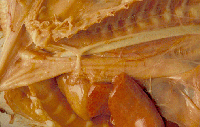
| + | [[Image:Praa.gif|thumb|right|200px|<center>Post-mortem specimen of an animal with a persistent right aortic arch <br><small> Alun Williams 2009 (RVC))<center></small>]] | ||
| + | This congenital condition is most commonly found in dogs after weaning, but is very rare in cats. | ||
| − | + | '''Persistent Right Aortic Arch''' is the most common vascular ring anomaly, increased incidence in German Shepherd Dogs, Irish setters and Great Danes. Vascular ring forms between the ductus arteriosus/ligamentum arteriosum and the persistent right aorta. [[Megaoesophagus]] is seen cranial to the constriction. Other causes include a '''double aortic arch''' and '''anomalous subclavian arteries'''. These arise from the aortic arch rather than the brachiocephalic artery. | |
| − | [[Megaoesophagus]] is seen cranial to the constriction. | ||
| − | + | During normal embryonic development there are five pairs of aortic arch arteries (1-6, 5 is absent) that undergo developmental changes necessary to form the major arteries of the head, neck, and upper thorax. | |
| − | |||
| − | + | Malformations of the aortic arch arteries can cause several vascular ring anomalies, but the most commonly seen anomaly is persistent right aortic arch (PRAA). With PRAA, the oesophagus and trachea are most often encircled by the left ligamentum arteriosum as they pass over the heart base causing chronic oesophageal and tracheal compression. | |
| − | |||
| − | + | When a young animal with PRAA is weaned onto solid food, the oesophageal and tracheal compression causes food stasis above the narrowed esophageal foramen leading to megaesophagus. Continued stress on the oesophagus in this manner can cause permanent damage. Additionally, [[Aspiration Pneumonia|aspiration pneumonia]] and the resulting respiratory distress is a complication of postprandial regurgitation. | |
| − | + | == Clinical Signs == | |
| − | |||
| − | |||
| − | '' | + | Clinical signs are usually due to constriction of the oesophagus, such as '''regurgitation of food''', usually noticed at weaning and aspiration pneumonia. This may be seen as '''coughing'''. |
| − | |||
| − | |||
| + | The animal may present as '[[vomiting]]' but thorough history and physical exam should distinguish this from regurgitation. | ||
| + | Respiratory distress may also be seen. '''Stunted growth''', thin and malnourished with an increased appetite may also be a clinical sign. | ||
| + | == Diagnosis == | ||
| + | History and clinical signs plus age and breed may be indicative of the disease. | ||
| − | + | On physical exam, there will be no cardiac murmurs unless a [[Patent Ductus Arteriosus]] (PDA) is also present. A palpable dilated cervical oesophagus may be detected. | |
| − | + | '''Radiographs''' should be taken for further diagnostics and will show a ventral deviation of the trachea and dilation of the oesophagus. A '''barium swallow''' will provide definitive diagnosis of this condition. | |
| − | + | == Treatment == | |
| − | + | '''Surgical resection''' should be performed as soon as possible. '''Aspiration pneumonia''' should be treated first if present, and dietary management should be put in place to minimise regurgitation: this involves feeding a liquid or gruel diet in small amounts from an elevated position. | |
| − | + | Surgical treatment involves '''transecting the ligamentum arteriosum''' via an intercostal thoracotomy. The ligament should be ligated before transection in case a [[Patent Ductus Arteriosus|patent ductus arteriosus]] is also present. | |
| − | + | A gastric tube is passed down the oesophagus to ensure no constrictions remain. | |
| − | |||
| − | |||
| − | |||
| − | == | + | == Prognosis == |
| − | + | '''Good with early surgical treatment''', but less favourable if there is extensive oesophageal damage. | |
| − | + | Post-operative care is aimed at preventing aspiration pneumonia, and mainly involves '''dietary management'''. Normal food can be gradually reintroduced after 4 weeks. | |
| − | + | If surgery cannot be performed, for whatever reason, then thoughts into the quality of life of the animal without surgery must be taken into consideration. | |
| − | = | + | {{Learning |
| − | = | + | |Vetstream = [https://www.vetstream.com/canis/Content/Disease/dis00916.asp, Vascular ring anomalies] |
| + | |flashcards = [[Small Animal Soft Tissue Surgery Q&A 19]] | ||
| − | + | [[Cardiovascular Developmental Pathology Flashcards]] | |
| + | }} | ||
| − | + | == References == | |
| − | + | Ettinger, S.J. and Feldman, E. C. (2000) '''Textbook of Veterinary Internal Medicine Diseases of the Dog and Cat '''Volume 2 (Fifth Edition)'' W.B. Saunders Company'' | |
| − | + | Ettinger, S.J, Feldman, E.C. (2005)''' Textbook of Veterinary Internal Medicine '''(6th edition, volume 2) ''W.B. Saunders Company'' | |
| − | + | Fossum, T. W. et. al. (2007) '''Small Animal Surgery''' (Third Edition) ''Mosby Elsevier '' | |
| − | + | Merck & Co (2008) '''The Merck Veterinary Manual '''(Eighth Edition) ''Merial'' | |
| − | + | Nelson, R.W. and Couto, C.G. (2009) '''Small Animal Internal Medicine '''(Fourth Edition) ''Mosby Elsevier'' | |
| − | |||
| − | + | {{review}} | |
| + | {{OpenPages}} | ||
| − | + | [[Category:Cardiovascular_System_-_Developmental_Pathology]] [[Category:Oesophagus_-_Pathology]] [[Category:Expert_Review - Small Animal]] [[Category:Cardiac_Diseases_-_Cat]] [[Category:Oesophageal_Diseases_-_Cat]] [[Category:Cardiac_Diseases_-_Dog]] [[Category:Oesophageal_Diseases_-_Dog]] | |
| − | + | [[Category:Cardiology Section]] | |
| − | |||
| − | |||
| − | |||
| − | |||
| − | |||
| − | |||
| − | |||
| − | |||
| − | |||
| − | |||
| − | |||
| − | |||
| − | |||
| − | |||
| − | |||
| − | |||
| − | |||
| − | |||
| − | |||
| − | |||
| − | |||
| − | |||
| − | |||
| − | |||
| − | [[ | ||
| − | |||
| − | |||
| − | |||
| − | |||
| − | |||
| − | |||
| − | |||
| − | |||
| − | [[ | ||
| − | |||
| − | |||
| − | |||
| − | [[Category: | ||
| − | [[Category: | ||
| − | [[Category: | ||
Latest revision as of 19:22, 1 September 2015
Conditions included: Persistent Right Aortic Arch (PRAA) — Double Aortic Arch — Anomalous Subclavian Arteries
Introduction
This congenital condition is most commonly found in dogs after weaning, but is very rare in cats.
Persistent Right Aortic Arch is the most common vascular ring anomaly, increased incidence in German Shepherd Dogs, Irish setters and Great Danes. Vascular ring forms between the ductus arteriosus/ligamentum arteriosum and the persistent right aorta. Megaoesophagus is seen cranial to the constriction. Other causes include a double aortic arch and anomalous subclavian arteries. These arise from the aortic arch rather than the brachiocephalic artery.
During normal embryonic development there are five pairs of aortic arch arteries (1-6, 5 is absent) that undergo developmental changes necessary to form the major arteries of the head, neck, and upper thorax.
Malformations of the aortic arch arteries can cause several vascular ring anomalies, but the most commonly seen anomaly is persistent right aortic arch (PRAA). With PRAA, the oesophagus and trachea are most often encircled by the left ligamentum arteriosum as they pass over the heart base causing chronic oesophageal and tracheal compression.
When a young animal with PRAA is weaned onto solid food, the oesophageal and tracheal compression causes food stasis above the narrowed esophageal foramen leading to megaesophagus. Continued stress on the oesophagus in this manner can cause permanent damage. Additionally, aspiration pneumonia and the resulting respiratory distress is a complication of postprandial regurgitation.
Clinical Signs
Clinical signs are usually due to constriction of the oesophagus, such as regurgitation of food, usually noticed at weaning and aspiration pneumonia. This may be seen as coughing.
The animal may present as 'vomiting' but thorough history and physical exam should distinguish this from regurgitation.
Respiratory distress may also be seen. Stunted growth, thin and malnourished with an increased appetite may also be a clinical sign.
Diagnosis
History and clinical signs plus age and breed may be indicative of the disease.
On physical exam, there will be no cardiac murmurs unless a Patent Ductus Arteriosus (PDA) is also present. A palpable dilated cervical oesophagus may be detected.
Radiographs should be taken for further diagnostics and will show a ventral deviation of the trachea and dilation of the oesophagus. A barium swallow will provide definitive diagnosis of this condition.
Treatment
Surgical resection should be performed as soon as possible. Aspiration pneumonia should be treated first if present, and dietary management should be put in place to minimise regurgitation: this involves feeding a liquid or gruel diet in small amounts from an elevated position.
Surgical treatment involves transecting the ligamentum arteriosum via an intercostal thoracotomy. The ligament should be ligated before transection in case a patent ductus arteriosus is also present.
A gastric tube is passed down the oesophagus to ensure no constrictions remain.
Prognosis
Good with early surgical treatment, but less favourable if there is extensive oesophageal damage.
Post-operative care is aimed at preventing aspiration pneumonia, and mainly involves dietary management. Normal food can be gradually reintroduced after 4 weeks.
If surgery cannot be performed, for whatever reason, then thoughts into the quality of life of the animal without surgery must be taken into consideration.
| Vascular Ring Anomalies Learning Resources | |
|---|---|
To reach the Vetstream content, please select |
Canis, Felis, Lapis or Equis |
 Test your knowledge using flashcard type questions |
Small Animal Soft Tissue Surgery Q&A 19 |
References
Ettinger, S.J. and Feldman, E. C. (2000) Textbook of Veterinary Internal Medicine Diseases of the Dog and Cat Volume 2 (Fifth Edition) W.B. Saunders Company
Ettinger, S.J, Feldman, E.C. (2005) Textbook of Veterinary Internal Medicine (6th edition, volume 2) W.B. Saunders Company
Fossum, T. W. et. al. (2007) Small Animal Surgery (Third Edition) Mosby Elsevier
Merck & Co (2008) The Merck Veterinary Manual (Eighth Edition) Merial
Nelson, R.W. and Couto, C.G. (2009) Small Animal Internal Medicine (Fourth Edition) Mosby Elsevier
| This article has been peer reviewed but is awaiting expert review. If you would like to help with this, please see more information about expert reviewing. |
Error in widget FBRecommend: unable to write file /var/www/wikivet.net/extensions/Widgets/compiled_templates/wrt69327f73997df7_84956632 Error in widget google+: unable to write file /var/www/wikivet.net/extensions/Widgets/compiled_templates/wrt69327f73a5cc18_85392209 Error in widget TwitterTweet: unable to write file /var/www/wikivet.net/extensions/Widgets/compiled_templates/wrt69327f73ae78e4_92240020
|
| WikiVet® Introduction - Help WikiVet - Report a Problem |
