Difference between revisions of "Heart Structure - Anatomy & Physiology"
Ggaitskell (talk | contribs) |
|||
| (19 intermediate revisions by 7 users not shown) | |||
| Line 1: | Line 1: | ||
| + | {{OpenPagesTop}} | ||
==Structure of the Heart== | ==Structure of the Heart== | ||
| − | + | [[Image:Aspinall Slide7.JPG|thumb|right|350px|<small>Image from [http://www.elsevierhealth.co.uk/veterinary-nursing/spe-60136/ Aspinall, The Complete Textbook of Veterinary Nursing], Elsevier Health Sciences, ''All rights reserved''</small>]] | |
| + | [[Image:Aspinall Slide8.JPG|thumb|right|350px|<small>Image from [http://www.elsevierhealth.co.uk/veterinary-nursing/spe-60136/ Aspinall, The Complete Textbook of Veterinary Nursing], Elsevier Health Sciences, ''All rights reserved''</small>]] | ||
===Position and Shape of the Heart=== | ===Position and Shape of the Heart=== | ||
| Line 13: | Line 15: | ||
===Pericardium=== | ===Pericardium=== | ||
| − | The pericardium is the membrane that surrounds and protects the heart. It is composed of two layers separated by a narrow cavity. The inner layer is firmly attached to the heart wall and is known as the visceral layer or epicardium. The outer layer is composed of relatively inelastic connective tissue and is termed the parietal layer. This fibrous layer prevents distension of the heart, thus preventing excessive stretching of the heart muscle fibres. The cavity between the two layers contains a small volume of fluid which serves as a lubricant, facilitating the movement of the heart by minimising friction. The sternopericardiac ligament connects the parietal layer to the sternum and the phrenopericardiac ligament joins the parietal layer to the diaphragm. The latter is present only in | + | The pericardium is the membrane that surrounds and protects the heart. It is composed of two layers separated by a narrow cavity. The inner layer is firmly attached to the heart wall and is known as the visceral layer or epicardium. The outer layer is composed of relatively inelastic connective tissue and is termed the parietal layer. This fibrous layer prevents distension of the heart, thus preventing excessive stretching of the heart muscle fibres. The cavity between the two layers contains a small volume of fluid which serves as a lubricant, facilitating the movement of the heart by minimising friction. The sternopericardiac ligament connects the parietal layer to the sternum and the phrenopericardiac ligament joins the parietal layer to the diaphragm. The latter is present only in canine and swine. |
===Layers of the Heart Wall=== | ===Layers of the Heart Wall=== | ||
| Line 43: | Line 45: | ||
The right atrium forms the dorsocranial section of the base of the heart and receives blood from the cranial vena cava, caudal vena cava and coronary sinus. The interatrial septum is a thin partition dividing the right and left atria and possesses a characteristic oval depression called the fossa ovalis which is a remnant of the foetal foramen ovalis. The right atrium also houses the sinoatrial node. Blood flows from the right atrium to the right ventricle through the tricuspid valve (also know as the right atrioventricular valve). | The right atrium forms the dorsocranial section of the base of the heart and receives blood from the cranial vena cava, caudal vena cava and coronary sinus. The interatrial septum is a thin partition dividing the right and left atria and possesses a characteristic oval depression called the fossa ovalis which is a remnant of the foetal foramen ovalis. The right atrium also houses the sinoatrial node. Blood flows from the right atrium to the right ventricle through the tricuspid valve (also know as the right atrioventricular valve). | ||
| + | In rabbits the right atrioventricular valve is bicuspid not tricuspid. | ||
====Right Ventricle==== | ====Right Ventricle==== | ||
| Line 57: | Line 60: | ||
<br> | <br> | ||
{{Template:Learning | {{Template:Learning | ||
| − | |dragster = [[Canine Heart Dissection Anatomy Resources (I & II)]]<br>[[Canine Heart Dissection Anatomy Resources (III & IV)]] | + | |dragster = [[Canine Heart Dissection Anatomy Resources (I & II)]]<br>[[Canine Heart Dissection Anatomy Resources (III & IV)]]<br>[[Cardiovascular System Histology Resource (I)]] |
| + | |videos = [[Video: Heart potcast|Heart potcast]]<br>[[Video: Heart (internal structure) potcast|Heart (internal structure) potcast]]<br>[[Video: Dorsal view of the ventricles and valves of the heart|Dorsal view of the ventricles and valves of the heart]] | ||
| + | |OVAM = [http://www.real3danatomy.com/thorax/dog-heart-lungs-ribs-3d.html Canine thoracic organs in Real 3D Anatomy, showing the heart, lungs and ribs and allowing detailed and accurate visualisation of organs "in-situ"]<br>[http://www.bristol.ac.uk/anatomy/media/elearning/internet/gracie/thorax/index.html Cross-sectional anatomy atlas of the canine thorax consisting of transverse sections and MRI images]<br>[http://www.um.es/anatvet/interactividad/ingles/avispi/practicas/practica3.htm Dissection plans of the thoracic cavity and heart]<br>[http://www.onlineveterinaryanatomy.net/sites/default/files/original_media/document/asset_8875_CDV.C.1_coronary_arteries_all.pdf Canine Heart - Coronary Artery Cast]<br>[http://www.onlineveterinaryanatomy.net/sites/default/files/original_media/document/asset_8864_cdv.c.18_all.pdf Plastinated Canine Heart (various views)]<br>[http://www.onlineveterinaryanatomy.net/content/cow-heart-internal-cast Internal Cast of Bovine Heart]<br>[http://www.onlineveterinaryanatomy.net/content/bronchial-and-heart-cast Canine Heart and Bronchial Cast] | ||
| + | |Vetstream = [https://www.vetstream.com/canis/Content/Disease/dis60195.asp Heart Failure] | ||
}} | }} | ||
| + | |||
| + | |||
| + | {{Chapter}} | ||
| + | {{Mansonchapter | ||
| + | |chapterlink = http://www.mansonpublishing.co.uk/book-images/9781840761535_sample.pdf | ||
| + | |chaptername = Normal Cardiovascular System | ||
| + | |book = Cardiovascular Disease in Small Animal Medicine | ||
| + | |author = Wendy A. Ware | ||
| + | |isbn = 9781840761535 | ||
| + | }} | ||
| + | |||
| + | == Webinars == | ||
| + | <rss max="10" highlight="heart cardiac cardiology">https://www.thewebinarvet.com/cardiology/webinars/feed</rss> | ||
[[Category:Heart - Anatomy & Physiology]] | [[Category:Heart - Anatomy & Physiology]] | ||
| + | [[Category:Cardiology Section]] | ||
Latest revision as of 19:27, 27 October 2022
Structure of the Heart


Position and Shape of the Heart
The heart is located in the thoracic cavity in between the lungs, 60% of it lying to the left of the median plane. The heart’s lateral projection extends from rib 3 to 6. Most of the heart’s surface is covered by the lungs and in juveniles it is bordered cranially by the thymus. Caudally the heart extends as far as the diaphragm. Variations in position and size exist among individuals depending on species, breed, age, fitness and pathology. Roughly speaking, the heart is responsible for about 0.75% of the bodyweight.
The heart is cone-shaped, with a broad base at the top from which the large blood vessels enter and exit. The tip, known as the apex, points downwards and lies close to the sternum. The longitudinal axis of the heart is tilted to varying degrees depending on the species resulting in the base facing craniodorsally and the apex caudoventrally.
The heart has a right and left lateral surface, which meet cranially at the right ventricular border and caudally at the left ventricular border. The auricles of the atria are visible on the left side, surrounding the root of the aorta and the pulmonary trunk, whilst the large veins and the main parts of the atria are situated on the right.
Grooves on the surface represent the divisions of the internal structure of the heart. The right surface of the heart is marked by the subsinusoidal groove which extends from the coronary groove to the apex of the heart. The paraconal groove runs over the left surface of the heart from the coronary groove to the distal end of the cranial margin. The fat-filled coronary groove contains the coronary blood vessels and marks the separation of the atria and ventricles.
Pericardium
The pericardium is the membrane that surrounds and protects the heart. It is composed of two layers separated by a narrow cavity. The inner layer is firmly attached to the heart wall and is known as the visceral layer or epicardium. The outer layer is composed of relatively inelastic connective tissue and is termed the parietal layer. This fibrous layer prevents distension of the heart, thus preventing excessive stretching of the heart muscle fibres. The cavity between the two layers contains a small volume of fluid which serves as a lubricant, facilitating the movement of the heart by minimising friction. The sternopericardiac ligament connects the parietal layer to the sternum and the phrenopericardiac ligament joins the parietal layer to the diaphragm. The latter is present only in canine and swine.
Layers of the Heart Wall
The wall of the heart consists of three layers: the epicardium (external layer), the myocardium (middle layer) and the endocardium (inner layer). The epicardium is the thin, transparent outer layer of the wall and is composed of delicate connective tissue. The myocardium, comprised of cardiac muscle tissue, makes up the majority of the cardiac wall and is responsible for its pumping action. The thickness of the myocardium mirrors the load to which each specific region of the heart is subjected. The endocardium is a thin layer of endothelium overlying a thin layer of connective tissue. It provides a smooth lining for the chambers of the heart and covers the valves. The endocardium is continuous with the endothelial lining of the large blood vessels attached to the heart.
Structure of Cardiac Muscle
Cardiac muscle fibres are shorter in length and larger in diameter than skeletal muscle fibres. They also exhibit branching, which gives an individual fibre a Y-shaped appearance. A typical cardiac muscle fibre is 50-100μm long and has a diameter of about 14μm. Normally, there is only one centrally located nucleus, although occasionally a cell may have two nuclei. The sarcoplasm of cardiac muscle is more abundant than that of skeletal muscle and the mitochondria are larger and more numerous. Cardiac muscle fibres have actin and myosin filaments arranged in the same way as skeletal muscle fibres and possess a well-developed T-tubule system. In contrast to skeletal muscle, cardiac muscle does not fatigue, cannot be repaired when damaged and is regulated by the autonomic nervous system.
Although cardiac muscle fibres branch and interconnect with each other, they form two separate functional syncytia, one for the atria and another for the ventricles. The ends of each fibre in a network connect to its neighbours by irregular transverse thickenings of the sarcolemma called intercalated discs. The discs contain desmosomes, which hold the fibres together, and gap junctions, which allow ions to travel between cells and permit the rapid propagation of action potentials. Consequently, excitement of a single fibre of either network results in stimulation of all the other fibres in the network. As a result, each network contracts as a functional unit.
Fibrous Skeleton
In addition to cardiac muscle tissue, the heart wall also contains dense connective tissue that forms the fibrous skeleton of the heart. The fibrous skeleton is composed of dense connective tissue rings that surround the four heart orifices. The skeleton contains fibrocartilage in which nodules of bones (ossa cordis) may develop in some species. Although these bones occur most commonly in cattle, they are not restricted to this species. The skeleton performs several functions:
- It serves as a point of attachment for the heart valves
- The cardiac muscle bundles insert onto the fibrous skeleton.
- It prevents the valves from overstretching as blood passes through them.
- It acts as an electrical insulator thereby preventing the direct spread of action potentials from the atria to the ventricles.
Chambers of the Heart
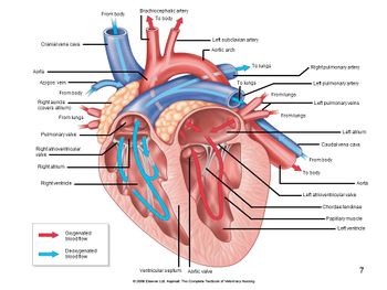
The heart contains four chambers. The two upper chambers are the atria and the two lower chambers are the ventricles. On the cranial surface of each atrium is a pouch-like appendage called an auricle which is thought to increase the capacity of the atrium slightly.
The thickness of the myocardium of the four chambers varies according to function. The atria are thin-walled because they deliver blood into the adjacent ventricles and the ventricles are equipped with thick muscular walls because they pump blood over greater distances. Even though the right and left ventricles act as two separate pumps that simultaneously eject equal volumes of blood, the right side has a much smaller workload. This is because the right ventricle only pumps blood into the lungs, which are close by and present little resistance to blood flow. On the other hand, the left ventricle pumps blood to the rest of the body, where the resistance to blood flow is considerably higher. Consequently, the left ventricle works harder than the right ventricle to maintain the same blood flow rate. This difference in workload affects the anatomy of the ventricular walls; the muscular wall of the left ventricle being significantly thicker than that of the right.
Right Atrium
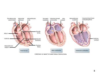
The right atrium forms the dorsocranial section of the base of the heart and receives blood from the cranial vena cava, caudal vena cava and coronary sinus. The interatrial septum is a thin partition dividing the right and left atria and possesses a characteristic oval depression called the fossa ovalis which is a remnant of the foetal foramen ovalis. The right atrium also houses the sinoatrial node. Blood flows from the right atrium to the right ventricle through the tricuspid valve (also know as the right atrioventricular valve). In rabbits the right atrioventricular valve is bicuspid not tricuspid.
Right Ventricle
The right ventricle forms most of the anterior surface of the heart and is crescent-shaped in cross-section. The cusps of the tricuspid valve are connected to tendon-like cords, the chordae tendinae, which, in turn, are connected to cone-shaped papillary muscles within the ventricular wall. The right ventricle is separated from the left by a partition called the interventricular septum. The trabecula septomarginalis is a muscular band that traverses the lumen of the right ventricle. Deoxygenated blood passes from the right ventricle through the pulmonary semi-lunar valve to the pulmonary trunk, which conveys the blood to the lungs.
Left Atrium
The left atrium forms the dorsocaudal section of the base of the heart and is similar to the right atrium in structure and shape. It receives oxygenated blood from the lungs via the pulmonary veins. Blood passes from the left atrium to the left ventricle through the bicuspid or left atrioventricular valve. The left atrium lies under the tracheal bifurcation and enlargement of this area of the heart can cause breathing difficulties.
Left Ventricle
The left ventricle forms the apex of the heart and is conical in shape. Blood passes from the left ventricle to the ascending aorta through the aortic semi-lunar valve. From here some of the blood flows into the coronary arteries, which branch from the ascending aorta and carry blood to the heart wall. The remainder of the blood travels throughout the body.
| Heart Structure - Anatomy & Physiology Learning Resources | |
|---|---|
To reach the Vetstream content, please select |
Canis, Felis, Lapis or Equis |
 Test your knowledge using drag and drop boxes |
Canine Heart Dissection Anatomy Resources (I & II) Canine Heart Dissection Anatomy Resources (III & IV) Cardiovascular System Histology Resource (I) |
 Selection of relevant videos |
Heart potcast Heart (internal structure) potcast Dorsal view of the ventricles and valves of the heart |
Anatomy Museum Resources |
Canine thoracic organs in Real 3D Anatomy, showing the heart, lungs and ribs and allowing detailed and accurate visualisation of organs "in-situ" Cross-sectional anatomy atlas of the canine thorax consisting of transverse sections and MRI images Dissection plans of the thoracic cavity and heart Canine Heart - Coronary Artery Cast Plastinated Canine Heart (various views) Internal Cast of Bovine Heart Canine Heart and Bronchial Cast |
| Sample Book Chapters | ||||
|---|---|---|---|---|
|
|
|
|
|
|
|
|
|
Webinars
Failed to load RSS feed from https://www.thewebinarvet.com/cardiology/webinars/feed: Error parsing XML for RSS