Difference between revisions of "Larynx - Anatomy & Physiology"
Fiorecastro (talk | contribs) |
|||
| (67 intermediate revisions by 7 users not shown) | |||
| Line 1: | Line 1: | ||
| − | {{ | + | {{OpenPagesTop}} |
| + | ==Introduction== | ||
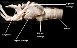
| + | [[Image:Larynx Anatomy.jpg|thumb|right|250px|Anatomy of the Larynx - Copyright University of Nottingham]] | ||
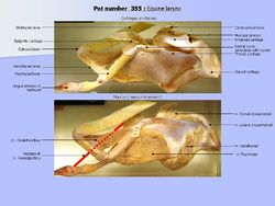
| + | [[Image: Annotated horse larynx.JPG|thumb|right|250px|Annotated horse larynx- Copyright RVC]] | ||
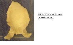
| + | [[Image:Epiglottic cartilage.jpg|thumb|right|250px|Epiglottic Cartilage - Copyright RVC]] | ||
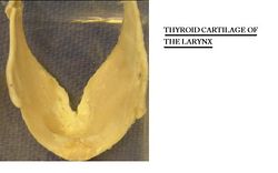
| + | [[Image:Thyroid cartilage.jpg|thumb|right|250px|Thyroid Cartilage - Copyright nabrown RVC]] | ||
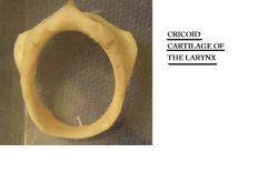
| + | [[Image:Cricoid cartilage.jpg|thumb|right|250px|Cricoid Cartilage - Copyright nabrown RVC]] | ||
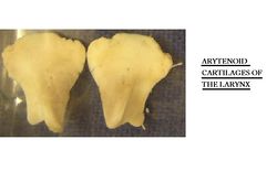
| + | [[Image:Arytenoid cartilages.jpg|thumb|right|250px|Arytenoid Cartilages - Copyright nabrown RVC]] | ||
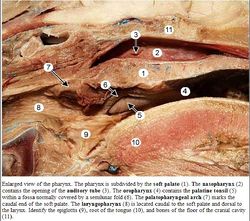
| + | [[Image:Pharynx Anatomy.jpg|thumb|right|250px|Pharynx Labelled - Copyright C.Clarkson and T.F.Fletcher University of Minnesota]] | ||
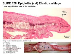
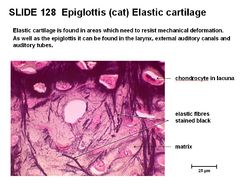
| + | [[Image:Epiglottis histology.jpg|right|thumb|250px|Epiglottis Histology - Copyright RVC]] | ||
| + | [[Image:Epiglottis histology 2.jpg|right|thumb|250px|Epiglottis Histology - Copyright RVC]] | ||
| − | + | The larynx is situated below where the [[Pharynx - Anatomy & Physiology|pharynx]] divides into the trachea and the [[Oesophagus - Anatomy & Physiology|oesophagus]]. It is contained partly within the rami of the mandible and extends caudally into the neck. '''Vocal folds''' and '''vestibular folds''' are present in the larynx and due to this, it is more commonly known as the voice box. | |
| − | |||
| − | |||
| − | |||
| − | |||
| − | |||
| − | | | ||
| − | | | ||
| − | |||
| − | |||
| − | |||
| − | The larynx | + | The cartilagenous larynx can be manually palpated in the living animal and is commonly implicated in respiratory conditions such as [[Laryngeal Paralysis|roaring]]. |
| − | |||
| − | |||
| − | |||
==Structure== | ==Structure== | ||
| − | + | The [[Pharynx - Anatomy & Physiology|pharynx]] is located rostrally to the larynx, whilst the trachea is located caudally. The larynx is suspended from the [[Hyoid Apparatus - Anatomy & Physiology|hyoid apparatus]]. It is bilaterally symmetrical and 'tube-shaped' and can be described as a '''musculocartilagenous organ'''. | |
| + | |||
| + | The larynx moves position when the animal [[Deglutition|swallows]] due to its attachments to the [[Tongue - Anatomy & Physiology|tongue]] and the '''basihyoid bone''' of the [[Hyoid Apparatus - Anatomy & Physiology|hyoid apparatus]] by the '''thyrohyoid membrane'''. | ||
| − | + | ===Synovial joints=== | |
| − | + | Synovial joints can be found between the '''thyrohyoid bone''' and the dorsorostral aspect of the '''thyroid cartilage'''. Synovial joints include the dorsal joint of '''thyroid cartilage'''; between the lateral aspect of the '''cricoid cartilage''' and the dorsocaudal aspect of the '''thyroid cartilage''' and between the '''cricoid''' and '''arytenoid cartilage'''. This allows abduction and adduction of the '''arytenoid cartilages'''. Movement of the '''cricoid-arytenoid joint''' controls the size of the glottic opening, the lumen and the larynx. | |
| − | + | ===Ligaments=== | |
| − | + | Membranes and elastic ligaments attach the laryngeal cartilages, allowing attachment of the epiglottis to the '''thyroid''' and '''cricoid cartilage'''. The first tracheal ring has attachment with the '''cricoid cartilage''' by the '''cricotracheal ligament'''. | |
| − | + | ===Extrinsic musculature=== | |
| − | + | Extrinsic musculature connects the larynx to the sternum, [[Tongue - Anatomy & Physiology|tongue]], [[Pharynx - Anatomy & Physiology|pharynx]] and [[Hyoid Apparatus - Anatomy & Physiology|hyoid apparatus]]. | |
| − | + | ===Intrinsic musculature=== | |
| − | |||
| − | |||
| − | |||
| − | |||
| − | + | Intrinsic musculature connects the laryngeal cartilages (see below). | |
| − | + | ===Vestibule=== | |
| − | |||
| − | |||
| − | + | The '''vestibule''' extends from the entrance of the larynx to the '''arytenoid cartilages''' and '''vocal folds'''. The '''vestibular folds''' run parallel, but rostral to, the '''vocal folds'''. | |
| − | + | ===Glottic cleft=== | |
| − | |||
| − | + | The '''glottic cleft''' (rima glottidis) is surrounded by the '''arytenoid cartilages''' dorsally and '''vocal cords''' ventrolaterally. It varies in size and is diamond shaped. The glottic cleft disappears when the glottis is closed. Vocal folds run caudodorsally. The '''infraglottic cavity''' extends from the caudal section of the '''arytenoid cartilages''' into the lumen of the trachea. It is fixed in size. | |
| − | |||
| − | + | ===Epiglottis=== | |
| − | |||
| − | |||
| − | |||
| − | |||
| − | + | The epiglottis is the rostral margin of the larynx. It is a flap of [[Cartilage - Anatomy & Physiology#Elastic Cartilage|elastic cartilage]] covered by mucous membrane. It forms the rostral boundary of the larynx and prevents food particles from entering the trachea. The epiglottis can return to its normal size and shape after distortion due to the vast amount of elastic fibres present within. | |
| − | |||
| − | + | Avian species do not have epiglottis. | |
==The Cartilage of the Larynx== | ==The Cartilage of the Larynx== | ||
===Thyroid Cartilage=== | ===Thyroid Cartilage=== | ||
| − | |||
| − | |||
| − | |||
| − | |||
| − | |||
| − | |||
| − | + | The thyroid cartilage is a [[Cartilage - Anatomy & Physiology#Hyaline Cartilage|hyaline cartilage]] and forms most of the floor of the larynx. The fusion of the two lateral plates varies in different species. The rostral part forms the 'Adam's apple'. The thyroid cartilage articulates with the '''thyrohyoid bone''' and the '''cricoid cartilage'''. It becomes brittle as the animal ages. | |
| − | |||
| − | |||
| − | |||
| − | |||
| − | |||
| − | |||
===Cricoid Cartilage=== | ===Cricoid Cartilage=== | ||
| − | |||
| − | |||
| − | |||
| − | |||
| − | |||
| − | |||
| − | + | The cricoid cartilage is also a [[Cartilage - Anatomy & Physiology#Hyaline Cartilage|hyaline cartilage]]. It is signet ring shaped and is wider on the dorsal surface than the ventral surface. There is a crest on the midline of the dorsal surface and facets for '''arytenoid cartilages''' on the rostral edge. The cricoid cartilage articulates with the '''thyroid cartilage'''. It also becomes brittle as the animal ages. | |
| − | |||
| − | |||
| − | |||
| − | |||
| − | |||
| − | |||
===Arytenoid Cartilage=== | ===Arytenoid Cartilage=== | ||
| − | |||
| − | |||
| − | + | The arytenoid cartilage is also a [[Cartilage - Anatomy & Physiology#Hyaline Cartilage|hyaline cartilage]]. It is paired and articulates with the rostral part of the '''cricoid cartilage'''. A '''vocal process''' is present on the caudal surface where the vocal folds attach; a muscular process extends laterally and a corniculate process extends dorsomedially. | |
| − | |||
| − | |||
| − | |||
| − | |||
| − | |||
| − | |||
===Epiglottic Cartilage=== | ===Epiglottic Cartilage=== | ||
| − | |||
| − | |||
| − | |||
| − | |||
| − | |||
| − | + | The epiglottic cartilage is an [[Cartilage - Anatomy & Physiology#Elastic Cartilage|elastic cartilage]], which is the most flexible and most rostral type of cartilage. The thinner stalk-like part, is attached to the root of the [[Tongue - Anatomy & Physiology|tongue]], the body of the '''thyroid cartilage''' and the '''basihyoid bone'''. The larger blade-like part lies behind the soft palate and points dorso-rostrally. During [[Deglutition|deglutition]], the large blade part of the '''epiglottic cartilage''' partially covers the entrance to the trachea. | |
| − | |||
| − | |||
| − | |||
| − | |||
| − | |||
| − | |||
===Interarytenoid Cartilage=== | ===Interarytenoid Cartilage=== | ||
| − | + | The interarytenoid cartilage is a nodule of [[Cartilage - Anatomy & Physiology#Hyaline Cartilage|hyaline cartilage]]. It is located between the '''arytenoid cartilages''' dorsally. | |
| − | |||
| − | |||
===Cuneiform Process=== | ===Cuneiform Process=== | ||
| − | + | The cuneiform process is formed by [[Cartilage - Anatomy & Physiology#Elastic Cartilage|elastic cartilage]]. It supports '''mucosal folds''' from the epiglottis to the '''arytenoid cartilages'''. It is not present in all species and can be free or fused with the '''epiglottic cartilages'''. | |
| − | |||
| − | |||
| − | |||
| − | |||
| − | |||
| − | |||
==The Vocal and Vestibular Folds== | ==The Vocal and Vestibular Folds== | ||
| Line 146: | Line 81: | ||
===Vocal Folds=== | ===Vocal Folds=== | ||
| − | + | The vocal folds are made of (slightly stiffer) elastic ligaments and pass between the '''arytenoid cartilages''' and the '''laryngeal floor'''. They run caudodorsally, with the ligament positioned medially and the '''vocalis muscle''' laterally. Fat surrounds the vocalis muscle. The vocal folds form part of the glottis and secrete mucous. They are used for vocalisation. | |
| − | |||
| − | |||
| − | |||
| − | |||
| − | |||
| − | |||
| − | |||
| − | |||
| − | |||
| − | |||
| − | |||
| − | |||
| − | |||
| − | |||
| − | |||
| − | |||
===Vestibular folds=== | ===Vestibular folds=== | ||
| − | + | The vestibular folds are made of (slightly stiffer) elastic ligaments. The '''vestibular ligaments''' are rostral to the '''vocal ligament'''. The vestibular folds run caudodorsally, rostral to the vocal folds with the ligament positioned medially and the '''vocalis muscle''' laterally. | |
| − | |||
| − | |||
| − | |||
| − | |||
| − | |||
| − | |||
| − | |||
| − | |||
| − | |||
| − | |||
| − | |||
| − | |||
| − | |||
| − | |||
==Intrinsic Musculature== | ==Intrinsic Musculature== | ||
| − | === | + | ===Cricothyroid muscle=== |
| − | + | The cricothyroid muscle is innervated by the '''cranial laryngeal nerve''', a branch of the vagus nerve ([[Cranial Nerves - Anatomy & Physiology|CN X]]). It moves the '''cricoid''' and '''arytenoid''' cartilages caudally to tense the vocal folds. | |
| − | |||
| − | |||
===Dorsal cricoarytenoid muscle=== | ===Dorsal cricoarytenoid muscle=== | ||
| − | + | The dorsal cricoarytenoid muscle is innervated by the '''caudal laryngeal nerve''', a branch of the vagus nerve ([[Cranial Nerves - Anatomy & Physiology|CN X]]). It runs from the dorsal surface of the '''cricoid cartilage''' to the '''arytenoid cartilage'''. It abducts the vocal process and therefore the vocal fold to widen the glottis. | |
| − | |||
| − | |||
| − | |||
===Lateral cricoarytenoid muscle=== | ===Lateral cricoarytenoid muscle=== | ||
| − | + | The lateral cricoarytenoid muscle is innervated by the '''caudal laryngeal nerve''', a branch of the vagus nerve ([[Cranial Nerves - Anatomy & Physiology|CN X]]). It adducts the vocal processes and narrows the glottis. | |
| − | |||
| − | |||
===Thyroarytenoid muscle=== | ===Thyroarytenoid muscle=== | ||
| − | + | The thyroarytenoid muscle is innervated by the '''caudal laryngeal nerve''', a branch of the vagus nerve ([[Cranial Nerves - Anatomy & Physiology|CN X]]). It runs from the laryngeal floor to the '''thyroid cartilage''' and '''arytenoid cartilage''' and alters the tension of the vocal and vestibular folds. It forms part of the '''sphincter muscular arrangement'''. | |
| − | |||
| − | |||
| − | |||
===Transverse arytenoid muscle=== | ===Transverse arytenoid muscle=== | ||
| − | + | The transverse arytenoid muscle is innervated by the '''caudal laryngeal nerve''', a branch of the vagus nerve ([[Cranial Nerves - Anatomy & Physiology|CN X]]). It completes the '''muscular sphincter arrangment''' and spans the '''arytenoid cartilages'''. | |
| − | |||
| − | |||
| − | |||
==Laryngeal Pharynx== | ==Laryngeal Pharynx== | ||
| − | + | The laryngeal pharynx is the largest part of the [[Pharynx - Anatomy & Physiology|pharynx]]. It is wide rostrally and narrows caudally. The laryngeal pharynx joins the [[Oesophagus - Anatomy & Physiology|oesophagus]] at the mucosal folds (clearest in the canine, in other species it is harder to see the demarcation). The lumen is closed at rest by the roof and walls falling towards the floor. The floor of the laryngeal pharynx contains the opening into the larynx - epiglottis, arytenoid cartilages and the aryepiglottic folds. | |
| − | |||
| − | |||
| − | |||
| − | |||
| − | |||
| − | |||
| − | |||
| − | |||
| − | |||
| − | |||
==Function== | ==Function== | ||
| − | + | The larynx protects the trachea in [[Deglutition|swallowing]], preventing aspiration of foreign material. During swallowing, the larynx is moved rostrally causing the epiglottis to partially cover the laryngeal entrance. Solid foods are carried over the laryngeal entrance by the muscles of the [[Pharynx - Anatomy & Physiology|pharynx]]. Fluids are deflected by the epiglottis. Closure of the glottis also prevents food passing down the larynx. The reflex stimulation of the mucosa promotes the coughing reflex. | |
| − | |||
| − | |||
| − | |||
| − | |||
| − | + | The larynx also allows the passage of air to the lungs and increases the intra-abdominal pressure. The glottis can widen by adduction of the vocal folds when breathing is vigorous. In addition to this, the larynx is used for vocalisation. | |
| − | + | ==Vasculature== | |
| − | + | '''Laryngeal branch of the superior thyroid artery''' supplies the rostral larynx and is a branch of the '''carotid artery'''. | |
| − | + | '''Laryngeal branch of inferior thyroid artery''' supplies the caudal larynx and is branching from the '''subclavian artery''' from the '''thyrocervical trunk'''. | |
| − | + | '''Laryngeal branch of cricothyroid artery''' branches from the '''superior thyroid artery'''. | |
| − | + | ==Innervation== | |
| − | |||
| − | == | ||
| − | |||
| − | |||
| − | |||
| − | |||
| − | |||
| − | |||
| − | |||
| − | |||
| − | + | The larynx is innervated by branches of the '''vagus nerve''' ([[Cranial Nerves - Anatomy & Physiology|CN X]]). | |
| − | |||
| − | + | '''Cranial laryngeal nerve''' has two branches. The internal branch innervates the mucosa and the external branch innervates the '''cricothyroid muscle''' and constricts the [[Pharynx - Anatomy & Physiology|pharynx]]. | |
| − | + | '''Caudal (recurrent) laryngeal nerve''' innervates the intrinsic muscles of the larynx (except the cricothyroid muscle). | |
| − | |||
| − | |||
| − | |||
| − | |||
| − | |||
==Lymphatics== | ==Lymphatics== | ||
| − | + | Lymphoid tissue is present. | |
| − | ==Species Differences== | + | ==[[Species Differences in Laryngeal Structure|Species Differences]]== |
| − | The variation between | + | The variation of the larynx between species is significant. More information can be found by clicking [[Species Differences in Laryngeal Structure|here]]. |
==Histology== | ==Histology== | ||
| − | + | Mucous glands are present in the larynx with a particularly high density in the ventricles. '''Stratified squamous epithelium''' is located rostrally around the laryngeal entrance, whilst '''ciliated pseudostratified columnar epithelium''' is elsewhere. | |
| − | |||
| − | |||
| − | + | The epiglottis is covered by mucous membrane and contains irregular elastic fibres. They form a dense network of branching fibres around the chondrocytes and less dense branching fibres towards the perichondrium. | |
| − | + | See [[Cartilage - Anatomy & Physiology#Elastic Cartilage|elastic cartilage histology]] for more information. | |
| − | |||
| − | |||
| − | |||
| − | |||
| + | See [[Cartilage - Anatomy & Physiology#Hyaline Cartilage|hyaline cartilage histology]] for more information. | ||
==Links== | ==Links== | ||
| − | [[Larynx - Pathology|Pathology of the Larynx]] | + | [[:Category:Larynx - Pathology|Pathology of the Larynx]] |
| − | [[ | + | [[Cartilage - Anatomy & Physiology|Cartilage - Anatomy & Physiology]] |
| − | [[ | + | [[Aspiration Pneumonia|Aspiration Pneumonia]] |
| − | [[Larynx - | + | {{Template:Learning |
| + | |flashcards = [[Facial_Muscles_-_Musculoskeletal_-_Flashcards|Facial Muscles]]<br>[[Pharynx_-_Musculoskeletal_-_Flashcards#The_Laryngeal_Pharynx_Flashcards|Larynx and Pharynx]]<br>[[Larynx_-_Musculoskeletal_-_Flashcards|Larynx]] | ||
| + | |powerpoints = [[Bone and Cartilage Histology resource|Interactive tutorial on bone and cartilage histology, including the Larynx]] | ||
| + | |videos = [http://stream2.rvc.ac.uk/Anatomy/canine/head_neck/Pot0258.mp4 Lateral section through the head of a dog] | ||
| + | |dragster = [[Canine Larynx Dissection Anatomy Resource]]<br>[[Canine Larynx Radiographical Anatomy Resource]] | ||
| + | |OVAM = [http://www.onlineveterinaryanatomy.net/sites/default/files/original_media/document/asset_9291_Dog%20and%20horse%20larynges%20PDF.pdf Comparative Larynges 1]<br>[http://www.onlineveterinaryanatomy.net/sites/default/files/original_media/document/asset_9289_Comparative%20larynges%20PDF.pdf Comparative Larynges 2]<br>[http://www.onlineveterinaryanatomy.net/content/dog-larynx-dorsal-view Canine Larynx - Dorsal View] | ||
| + | }} | ||
| − | + | {{review}} | |
| + | ==Webinars== | ||
| + | <rss max="10" highlight="none">https://www.thewebinarvet.com/respiratory/webinars/feed</rss> | ||
| − | [ | + | [[Category:Respiratory System - Anatomy & Physiology]][[Category:Musculoskeletal System - Anatomy & Physiology]] |
| + | [[Category:A&P Done]] | ||
Latest revision as of 17:41, 7 November 2022
Introduction
The larynx is situated below where the pharynx divides into the trachea and the oesophagus. It is contained partly within the rami of the mandible and extends caudally into the neck. Vocal folds and vestibular folds are present in the larynx and due to this, it is more commonly known as the voice box.
The cartilagenous larynx can be manually palpated in the living animal and is commonly implicated in respiratory conditions such as roaring.
Structure
The pharynx is located rostrally to the larynx, whilst the trachea is located caudally. The larynx is suspended from the hyoid apparatus. It is bilaterally symmetrical and 'tube-shaped' and can be described as a musculocartilagenous organ.
The larynx moves position when the animal swallows due to its attachments to the tongue and the basihyoid bone of the hyoid apparatus by the thyrohyoid membrane.
Synovial joints
Synovial joints can be found between the thyrohyoid bone and the dorsorostral aspect of the thyroid cartilage. Synovial joints include the dorsal joint of thyroid cartilage; between the lateral aspect of the cricoid cartilage and the dorsocaudal aspect of the thyroid cartilage and between the cricoid and arytenoid cartilage. This allows abduction and adduction of the arytenoid cartilages. Movement of the cricoid-arytenoid joint controls the size of the glottic opening, the lumen and the larynx.
Ligaments
Membranes and elastic ligaments attach the laryngeal cartilages, allowing attachment of the epiglottis to the thyroid and cricoid cartilage. The first tracheal ring has attachment with the cricoid cartilage by the cricotracheal ligament.
Extrinsic musculature
Extrinsic musculature connects the larynx to the sternum, tongue, pharynx and hyoid apparatus.
Intrinsic musculature
Intrinsic musculature connects the laryngeal cartilages (see below).
Vestibule
The vestibule extends from the entrance of the larynx to the arytenoid cartilages and vocal folds. The vestibular folds run parallel, but rostral to, the vocal folds.
Glottic cleft
The glottic cleft (rima glottidis) is surrounded by the arytenoid cartilages dorsally and vocal cords ventrolaterally. It varies in size and is diamond shaped. The glottic cleft disappears when the glottis is closed. Vocal folds run caudodorsally. The infraglottic cavity extends from the caudal section of the arytenoid cartilages into the lumen of the trachea. It is fixed in size.
Epiglottis
The epiglottis is the rostral margin of the larynx. It is a flap of elastic cartilage covered by mucous membrane. It forms the rostral boundary of the larynx and prevents food particles from entering the trachea. The epiglottis can return to its normal size and shape after distortion due to the vast amount of elastic fibres present within.
Avian species do not have epiglottis.
The Cartilage of the Larynx
Thyroid Cartilage
The thyroid cartilage is a hyaline cartilage and forms most of the floor of the larynx. The fusion of the two lateral plates varies in different species. The rostral part forms the 'Adam's apple'. The thyroid cartilage articulates with the thyrohyoid bone and the cricoid cartilage. It becomes brittle as the animal ages.
Cricoid Cartilage
The cricoid cartilage is also a hyaline cartilage. It is signet ring shaped and is wider on the dorsal surface than the ventral surface. There is a crest on the midline of the dorsal surface and facets for arytenoid cartilages on the rostral edge. The cricoid cartilage articulates with the thyroid cartilage. It also becomes brittle as the animal ages.
Arytenoid Cartilage
The arytenoid cartilage is also a hyaline cartilage. It is paired and articulates with the rostral part of the cricoid cartilage. A vocal process is present on the caudal surface where the vocal folds attach; a muscular process extends laterally and a corniculate process extends dorsomedially.
Epiglottic Cartilage
The epiglottic cartilage is an elastic cartilage, which is the most flexible and most rostral type of cartilage. The thinner stalk-like part, is attached to the root of the tongue, the body of the thyroid cartilage and the basihyoid bone. The larger blade-like part lies behind the soft palate and points dorso-rostrally. During deglutition, the large blade part of the epiglottic cartilage partially covers the entrance to the trachea.
Interarytenoid Cartilage
The interarytenoid cartilage is a nodule of hyaline cartilage. It is located between the arytenoid cartilages dorsally.
Cuneiform Process
The cuneiform process is formed by elastic cartilage. It supports mucosal folds from the epiglottis to the arytenoid cartilages. It is not present in all species and can be free or fused with the epiglottic cartilages.
The Vocal and Vestibular Folds
Vocal Folds
The vocal folds are made of (slightly stiffer) elastic ligaments and pass between the arytenoid cartilages and the laryngeal floor. They run caudodorsally, with the ligament positioned medially and the vocalis muscle laterally. Fat surrounds the vocalis muscle. The vocal folds form part of the glottis and secrete mucous. They are used for vocalisation.
Vestibular folds
The vestibular folds are made of (slightly stiffer) elastic ligaments. The vestibular ligaments are rostral to the vocal ligament. The vestibular folds run caudodorsally, rostral to the vocal folds with the ligament positioned medially and the vocalis muscle laterally.
Intrinsic Musculature
Cricothyroid muscle
The cricothyroid muscle is innervated by the cranial laryngeal nerve, a branch of the vagus nerve (CN X). It moves the cricoid and arytenoid cartilages caudally to tense the vocal folds.
Dorsal cricoarytenoid muscle
The dorsal cricoarytenoid muscle is innervated by the caudal laryngeal nerve, a branch of the vagus nerve (CN X). It runs from the dorsal surface of the cricoid cartilage to the arytenoid cartilage. It abducts the vocal process and therefore the vocal fold to widen the glottis.
Lateral cricoarytenoid muscle
The lateral cricoarytenoid muscle is innervated by the caudal laryngeal nerve, a branch of the vagus nerve (CN X). It adducts the vocal processes and narrows the glottis.
Thyroarytenoid muscle
The thyroarytenoid muscle is innervated by the caudal laryngeal nerve, a branch of the vagus nerve (CN X). It runs from the laryngeal floor to the thyroid cartilage and arytenoid cartilage and alters the tension of the vocal and vestibular folds. It forms part of the sphincter muscular arrangement.
Transverse arytenoid muscle
The transverse arytenoid muscle is innervated by the caudal laryngeal nerve, a branch of the vagus nerve (CN X). It completes the muscular sphincter arrangment and spans the arytenoid cartilages.
Laryngeal Pharynx
The laryngeal pharynx is the largest part of the pharynx. It is wide rostrally and narrows caudally. The laryngeal pharynx joins the oesophagus at the mucosal folds (clearest in the canine, in other species it is harder to see the demarcation). The lumen is closed at rest by the roof and walls falling towards the floor. The floor of the laryngeal pharynx contains the opening into the larynx - epiglottis, arytenoid cartilages and the aryepiglottic folds.
Function
The larynx protects the trachea in swallowing, preventing aspiration of foreign material. During swallowing, the larynx is moved rostrally causing the epiglottis to partially cover the laryngeal entrance. Solid foods are carried over the laryngeal entrance by the muscles of the pharynx. Fluids are deflected by the epiglottis. Closure of the glottis also prevents food passing down the larynx. The reflex stimulation of the mucosa promotes the coughing reflex.
The larynx also allows the passage of air to the lungs and increases the intra-abdominal pressure. The glottis can widen by adduction of the vocal folds when breathing is vigorous. In addition to this, the larynx is used for vocalisation.
Vasculature
Laryngeal branch of the superior thyroid artery supplies the rostral larynx and is a branch of the carotid artery.
Laryngeal branch of inferior thyroid artery supplies the caudal larynx and is branching from the subclavian artery from the thyrocervical trunk.
Laryngeal branch of cricothyroid artery branches from the superior thyroid artery.
Innervation
The larynx is innervated by branches of the vagus nerve (CN X).
Cranial laryngeal nerve has two branches. The internal branch innervates the mucosa and the external branch innervates the cricothyroid muscle and constricts the pharynx.
Caudal (recurrent) laryngeal nerve innervates the intrinsic muscles of the larynx (except the cricothyroid muscle).
Lymphatics
Lymphoid tissue is present.
Species Differences
The variation of the larynx between species is significant. More information can be found by clicking here.
Histology
Mucous glands are present in the larynx with a particularly high density in the ventricles. Stratified squamous epithelium is located rostrally around the laryngeal entrance, whilst ciliated pseudostratified columnar epithelium is elsewhere.
The epiglottis is covered by mucous membrane and contains irregular elastic fibres. They form a dense network of branching fibres around the chondrocytes and less dense branching fibres towards the perichondrium.
See elastic cartilage histology for more information.
See hyaline cartilage histology for more information.
Links
Cartilage - Anatomy & Physiology
| Larynx - Anatomy & Physiology Learning Resources | |
|---|---|
 Test your knowledge using drag and drop boxes |
Canine Larynx Dissection Anatomy Resource Canine Larynx Radiographical Anatomy Resource |
 Test your knowledge using flashcard type questions |
Facial Muscles Larynx and Pharynx Larynx |
 Selection of relevant videos |
Lateral section through the head of a dog |
 Selection of relevant PowerPoint tutorials |
Interactive tutorial on bone and cartilage histology, including the Larynx |
Anatomy Museum Resources |
Comparative Larynges 1 Comparative Larynges 2 Canine Larynx - Dorsal View |
| This article has been peer reviewed but is awaiting expert review. If you would like to help with this, please see more information about expert reviewing. |
Webinars
Failed to load RSS feed from https://www.thewebinarvet.com/respiratory/webinars/feed: Error parsing XML for RSS
