Difference between revisions of "Mammary Gland - Anatomy & Physiology"
Fiorecastro (talk | contribs) |
|||
| (131 intermediate revisions by 10 users not shown) | |||
| Line 1: | Line 1: | ||
| − | + | {{OpenPagesTop}} | |
| + | == Introduction == | ||
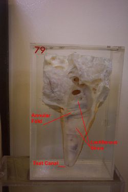
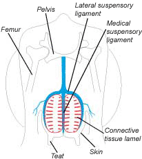
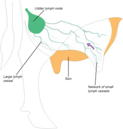
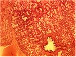
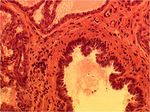
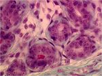
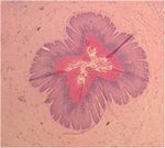
| − | + | [[Image:Cow teat 4.jpg|thumb|right|250px|Dissection of a Teat of the Bovine Udder,Courtesy of Andrew Crook, Copyright RVC 2008]] [[Image:Suspensory structure of udder.gif|thumb|right|250px|Suspensory apparatus of the udder]] [[Image:Udder lymphatics.gif|thumb|right|250px|Lymphatic drainage of the udder, Copyright DeLaval 2008]] [[Image:Active Mammary Gland.jpg|thumb|right|150px|<small><center> The Active Mammary Gland. Copyright RVC 2008 (Courtesy of Tanya Hopcroft (RVC))</center></small>]] [[Image:Active Mammary Gland high power.jpg|thumb|right|150px|<small><center> The Active Mammary Gland at High Power. Copyright RVC 2008 (Courtesy of Tanya Hopcroft (RVC))</center></small>]] [[Image:Mammary Gland myoepithelial cells.jpg|thumb|right|150px|<small><center> Mammary Myoepithelial Cells. Copyright RVC 2008 (Courtesy of Tanya Hopcroft (RVC))</center></small>]] [[Image:Mammary Gland lactiferous duct.jpg|thumb|right|150px|<small><center> Section of the Mammary Gland showing a Lactiferous Duct. Copyright RVC 2008 (Courtesy of Tanya Hopcroft (RVC))</center></small>]] [[Image:Mammary Gland teat canal keratin plug.jpg|thumb|right|150px|<small><center> Cross Section through the Teat Canal of the Mammary Gland showing a Keratin Plug. Copyright RVC 2008 (Courtesy of Tanya Hopcroft (RVC))</center></small>]] | |
| − | |||
| − | |||
| − | |||
| − | |||
| + | The mammary gland is a modified sweat gland that nourishes the young. It consists of the '''mamma''' and the '''teat'''. Undeveloped in both the male and female at birth, the female mammary gland begins to develop as a secondary sex characteristic at puberty. With the birth of the first young, and first lactation, the mammary gland attains its full size and function. When suckling by the young stops, milk production ceases and the gland regresses. Shortly before the next and subsequent parturitions, the gland is stimulated by hormonal changes to produce milk. | ||
| − | + | == Development of the Mammary Gland (prenatal mammogenesis) == | |
| − | + | An ectodermal thickening developes along the ventral body wall extending from the thoracic to inguinal region - this is the '''mammary ridge'''. Cells aggregate, multiply and differentiate to form a chain of condensed '''mammary buds'''. Most mammary buds regress but those that remain each develops into a '''mammary gland'''. A mammary gland is the secretory and duct system associated with one teat. Mammary buds grow into overlying mesenchyme, and '''primary epidermal sprouts''' grow out of the bud apex. The epidermal sprout branches extensively and develops a complete '''duct system'''. Mammary adipose tissue is derived from mesoderm. This is required for complete mammary development and is absent in the male. As a result, mammary development in the male is halted at the epidermal sprout stage. | |
| − | |||
| − | |||
| − | |||
| − | |||
| − | |||
| − | |||
| + | == Structure == | ||
| − | ''''' | + | The '''mamma (pleural = mammae) ''' is the glandular structure associated with a '''papilla '''(teat) and may contain one or more duct systems. The '''udder''' is a term designating all the mammae in the ruminant and the mare (sometimes also used for the sow). The '''lobes''' are the internal compartments of the mamma, separated by adipose tissue. The lobes are divided into '''lobules''', consisting of connective tissue containing '''alveoli''', which are clusters of milk secreting cells. The '''lactiferous ducts''' are large ducts conveying milk from the alveoli to the '''lactiferous sinus'''. The openings of the lactiferous ducts convey milk formed in the alveolus to the gland sinus. |
| − | '' | + | The '''lactiferous sinus''' (milk sinus) is the milk storage cavity within the teat and glandular body. The '''gland sinus''' is part of the milk sinus within the glandular body and the '''teat sinus''' is part of the milk sinus within the teat. |
| − | + | The '''teat''' is the projecting part of the mammary gland containing part of the milk sinus. The '''papillary duct '''(teat canal) is the canal leading from the teat sinus to the teat opening and may be single or multiple. The '''ostium '''(teat opening) is the opening of the papillary duct and the exit point for milk or entrance point for bacteria. The '''sphincter''' consists of muscular fibres surrounding the teat opening that prevent milk flow except during suckling or milking. | |
| − | |||
| − | |||
| − | + | === Suspensory Apparatus === | |
| − | |||
| + | In species with large udders, especially in dairy cattle, there is a suspensory apparatus, which is organised into the lateral and medial laminae suspending the mammary gland from the ventral aspect of the trunk by their attachment to the pubic symphysis. The '''lateral lamina''' consists of collagen fibres from the fascia of the pubic symphysis and the edge of the superficial inguinal ring. The '''medial lamina''' consists of elastic fibres from the tunica flava ventral to the pubic symphysis The '''intermammary groove''' divides the left and right rows of mammae. | ||
| + | == Blood Supply == | ||
| − | + | === Arteries === | |
| − | |||
| − | |||
| + | The main blood supply to the inguinal mammary glands is from the '''external pudendal artery'''. This arises indirectly from the external iliac artery via the deep femoral artery. The external pudendal artery passes through the inguinal canal. In species which also have '''thoracic and abdominal mammary glands''' (bitch, queen, sow) additional blood supply is derived from the '''internal thoracic artery''' and its branches - cranial superficial epigastric arteries as well as from '''lateral thoracic''' and '''intercostal arteries'''. | ||
| + | === Veins === | ||
| − | '' | + | In most species'''thoracic and cranial abdominal mammary glands '''drain via '''cranial superficial epigastric veins''' into the '''internal thoracic vein'''. '''Caudal abdominal and inguinal mammary glands '''drain via '''caudal superficial epigastric veins''' into the '''external pudendal vein'''. |
| − | |||
| − | |||
| − | |||
| + | In cattle a venous ring is formed between the base of the udder and the abdominal wall. During the first pregnancy, an anastamosis develops between cranial and caudal superficial epigastric veins forming the '''subcutaneous abdominal vein''' (milk vein). As a result some drainage from venous ring passes in a cranial direction via this vessel, which then drains deeply through the abdominal wall (milk well) into the internal thoracic vein. Other drainage passes to the external pudendal veins or to perineal veins.<br> | ||
| + | == Innervation == | ||
| − | '' | + | Somatic innervation is via the ventral rami of the spinal nerves. In the cow, the ventral branches of L1 and L2 ('''iliohypogastric and ilioinguinal''') supply the skin of the cranial glands. Mammary branches of the '''pudendal nerve''' supply the caudal aspect of the udder. There is sympathetic innervation to the blood vessels and teat sphincter smooth muscle. Mammary glands also have major influence from endocrine hormones. |
| − | + | == Lymphatics == | |
| − | |||
| − | |||
| + | The more caudal mammary glands drain to the '''superficial inguinal lymph node''' and the more cranial mammary glands to the axillary or sternal lymph nodes. | ||
| + | === Lymphatic drainage in the cow === | ||
| − | ''''' | + | The '''afferent lymphatic ducts''' pass dorsocaudally to reach the '''superficial inguinal''' (mammary) lymph nodes at the dorsocaudal side of the udder. These are usually palpable large, kidney-shaped nodes between the caudal side of the udder base and the thigh. The '''efferent lymphatic ducts''' pass into the abdomen through the inguinal canal to empty into the deep inguinal node. |
| − | + | == Histology == | |
| − | |||
| − | |||
| − | |||
| − | [[ | + | Secretory tissue is arranged into '''lobes''', each consisting of many '''lobules'''. Each lobule contains groups of '''alveoli''' (secretory compound tubuloalveolar cells) surrounded by a network of blood vessels and connective tissue stroma. The alveolar lumen is filled with milk during lactation. '''Myoepithelial cells''' lie between alveolar epithelial cells and the basement membrane. These contract under the influence of oxytocin to release milk to the exterior. Lobes and lobules are drained by lactiferous ducts into the '''gland sinus''', which is continuous with the '''teat sinus'''. The epithelium lining the lactiferous ducts and the sinus is two-layered cuboidal. A '''teat canal''' connects the teat sinus to the exterior. The lining is stratified squamous epithelium. Circular smooth muscle in the wall of the canal forms a '''sphincter'''. Between milkings, the narrow lumen of the teat canal is filled with a soft keratin plug to prevent bacteria entering the teat sinus and prevent milk leakage. |
| + | |||
| + | == Species Differences == | ||
| + | |||
| + | '''Position and Morphology''' | ||
| + | |||
| + | <br> | ||
| + | |||
| + | {| border="1" style="width: 75%; height: 200px;" | ||
| + | |- | ||
| + | ! Species | ||
| + | ! '''Primates''' | ||
| + | ! '''Elephant''' | ||
| + | ! '''Goat and Sheep''' | ||
| + | ! '''Guinea Pig''' | ||
| + | ! '''Cow''' | ||
| + | ! '''Mare''' | ||
| + | ! '''Rat''' | ||
| + | ! '''Dog''' | ||
| + | ! '''Sow''' | ||
| + | ! '''Cat''' | ||
| + | |- | ||
| + | | '''Number of Mammae/teats''' | ||
| + | | 2 | ||
| + | | 2 | ||
| + | | 2 | ||
| + | | 2 | ||
| + | | 4 | ||
| + | | 4 (2 teats) | ||
| + | | ~10 | ||
| + | | ~10 | ||
| + | | 8-18 | ||
| + | | 8 | ||
| + | |- | ||
| + | | '''Position''' | ||
| + | | Pectoral | ||
| + | | Pectoral | ||
| + | | Inguinal | ||
| + | | Inguinal | ||
| + | | Inguinal | ||
| + | | Inguinal | ||
| + | | Abdominal,Ventral | ||
| + | | Thoracoabdominoinguinal | ||
| + | | Thoracoabdominoinguinal | ||
| + | | Thoracoabdominal | ||
| + | |- | ||
| + | | '''Teat Ducts''' | ||
| + | | 10-20 | ||
| + | | Several | ||
| + | | 1 | ||
| + | | 1 | ||
| + | | 1 | ||
| + | | 2 | ||
| + | | 1 | ||
| + | | 8-22 | ||
| + | | 2 | ||
| + | | 4-8 | ||
| + | |} | ||
| + | |||
| + | <br> Click here for more information on the [[Cow Mammary Gland - Anatomy & Physiology|cow]], [[Small Ruminant Mammary Gland - Anatomy & Physiology|small ruminant]], [[Sow Mammary Gland - Anatomy & Physiology|sow]], [[Mare Mammary Gland - Anatomy & Physiology|mare]] and [[Carnivore Mammary Gland - Anatomy & Physiology|carnivore]]. | ||
| + | |||
| + | {{Template:Learning | ||
| + | |flashcards = [[Reproductive System Flashcards - Anatomy & Physiology|Mammary gland flashcards]] | ||
| + | |powerpoints = [[Mammary Gland Histology resource|Histology of the mammary gland]] | ||
| + | |videos = [[Video: Bovine teat and mammary gland potcast|Bovine teat and mammary gland potcast]]<br>[[Video: Mammary gland of the ewe|Mammary gland of the ewe]] | ||
| + | }} | ||
| + | <br> | ||
| + | {{Template:David Hogg reviewed | ||
| + | |date=20/03/2011}} | ||
| + | |||
| + | |||
| + | ==Webinars== | ||
| + | <rss max="10" highlight="none">https://www.thewebinarvet.com/urogenital-and-reprodcution/webinars/feed</rss> | ||
| + | |||
| + | [[Category:Female_Reproduction]] [[Category:Integumentary_System_-_Anatomy_&_Physiology]] [[Category:A&P_Done]] [[Category:David_Hogg_reviewed]] | ||
Latest revision as of 17:16, 7 December 2022
Introduction
The mammary gland is a modified sweat gland that nourishes the young. It consists of the mamma and the teat. Undeveloped in both the male and female at birth, the female mammary gland begins to develop as a secondary sex characteristic at puberty. With the birth of the first young, and first lactation, the mammary gland attains its full size and function. When suckling by the young stops, milk production ceases and the gland regresses. Shortly before the next and subsequent parturitions, the gland is stimulated by hormonal changes to produce milk.
Development of the Mammary Gland (prenatal mammogenesis)
An ectodermal thickening developes along the ventral body wall extending from the thoracic to inguinal region - this is the mammary ridge. Cells aggregate, multiply and differentiate to form a chain of condensed mammary buds. Most mammary buds regress but those that remain each develops into a mammary gland. A mammary gland is the secretory and duct system associated with one teat. Mammary buds grow into overlying mesenchyme, and primary epidermal sprouts grow out of the bud apex. The epidermal sprout branches extensively and develops a complete duct system. Mammary adipose tissue is derived from mesoderm. This is required for complete mammary development and is absent in the male. As a result, mammary development in the male is halted at the epidermal sprout stage.
Structure
The mamma (pleural = mammae) is the glandular structure associated with a papilla (teat) and may contain one or more duct systems. The udder is a term designating all the mammae in the ruminant and the mare (sometimes also used for the sow). The lobes are the internal compartments of the mamma, separated by adipose tissue. The lobes are divided into lobules, consisting of connective tissue containing alveoli, which are clusters of milk secreting cells. The lactiferous ducts are large ducts conveying milk from the alveoli to the lactiferous sinus. The openings of the lactiferous ducts convey milk formed in the alveolus to the gland sinus.
The lactiferous sinus (milk sinus) is the milk storage cavity within the teat and glandular body. The gland sinus is part of the milk sinus within the glandular body and the teat sinus is part of the milk sinus within the teat.
The teat is the projecting part of the mammary gland containing part of the milk sinus. The papillary duct (teat canal) is the canal leading from the teat sinus to the teat opening and may be single or multiple. The ostium (teat opening) is the opening of the papillary duct and the exit point for milk or entrance point for bacteria. The sphincter consists of muscular fibres surrounding the teat opening that prevent milk flow except during suckling or milking.
Suspensory Apparatus
In species with large udders, especially in dairy cattle, there is a suspensory apparatus, which is organised into the lateral and medial laminae suspending the mammary gland from the ventral aspect of the trunk by their attachment to the pubic symphysis. The lateral lamina consists of collagen fibres from the fascia of the pubic symphysis and the edge of the superficial inguinal ring. The medial lamina consists of elastic fibres from the tunica flava ventral to the pubic symphysis The intermammary groove divides the left and right rows of mammae.
Blood Supply
Arteries
The main blood supply to the inguinal mammary glands is from the external pudendal artery. This arises indirectly from the external iliac artery via the deep femoral artery. The external pudendal artery passes through the inguinal canal. In species which also have thoracic and abdominal mammary glands (bitch, queen, sow) additional blood supply is derived from the internal thoracic artery and its branches - cranial superficial epigastric arteries as well as from lateral thoracic and intercostal arteries.
Veins
In most speciesthoracic and cranial abdominal mammary glands drain via cranial superficial epigastric veins into the internal thoracic vein. Caudal abdominal and inguinal mammary glands drain via caudal superficial epigastric veins into the external pudendal vein.
In cattle a venous ring is formed between the base of the udder and the abdominal wall. During the first pregnancy, an anastamosis develops between cranial and caudal superficial epigastric veins forming the subcutaneous abdominal vein (milk vein). As a result some drainage from venous ring passes in a cranial direction via this vessel, which then drains deeply through the abdominal wall (milk well) into the internal thoracic vein. Other drainage passes to the external pudendal veins or to perineal veins.
Innervation
Somatic innervation is via the ventral rami of the spinal nerves. In the cow, the ventral branches of L1 and L2 (iliohypogastric and ilioinguinal) supply the skin of the cranial glands. Mammary branches of the pudendal nerve supply the caudal aspect of the udder. There is sympathetic innervation to the blood vessels and teat sphincter smooth muscle. Mammary glands also have major influence from endocrine hormones.
Lymphatics
The more caudal mammary glands drain to the superficial inguinal lymph node and the more cranial mammary glands to the axillary or sternal lymph nodes.
Lymphatic drainage in the cow
The afferent lymphatic ducts pass dorsocaudally to reach the superficial inguinal (mammary) lymph nodes at the dorsocaudal side of the udder. These are usually palpable large, kidney-shaped nodes between the caudal side of the udder base and the thigh. The efferent lymphatic ducts pass into the abdomen through the inguinal canal to empty into the deep inguinal node.
Histology
Secretory tissue is arranged into lobes, each consisting of many lobules. Each lobule contains groups of alveoli (secretory compound tubuloalveolar cells) surrounded by a network of blood vessels and connective tissue stroma. The alveolar lumen is filled with milk during lactation. Myoepithelial cells lie between alveolar epithelial cells and the basement membrane. These contract under the influence of oxytocin to release milk to the exterior. Lobes and lobules are drained by lactiferous ducts into the gland sinus, which is continuous with the teat sinus. The epithelium lining the lactiferous ducts and the sinus is two-layered cuboidal. A teat canal connects the teat sinus to the exterior. The lining is stratified squamous epithelium. Circular smooth muscle in the wall of the canal forms a sphincter. Between milkings, the narrow lumen of the teat canal is filled with a soft keratin plug to prevent bacteria entering the teat sinus and prevent milk leakage.
Species Differences
Position and Morphology
| Species | Primates | Elephant | Goat and Sheep | Guinea Pig | Cow | Mare | Rat | Dog | Sow | Cat |
|---|---|---|---|---|---|---|---|---|---|---|
| Number of Mammae/teats | 2 | 2 | 2 | 2 | 4 | 4 (2 teats) | ~10 | ~10 | 8-18 | 8 |
| Position | Pectoral | Pectoral | Inguinal | Inguinal | Inguinal | Inguinal | Abdominal,Ventral | Thoracoabdominoinguinal | Thoracoabdominoinguinal | Thoracoabdominal |
| Teat Ducts | 10-20 | Several | 1 | 1 | 1 | 2 | 1 | 8-22 | 2 | 4-8 |
Click here for more information on the cow, small ruminant, sow, mare and carnivore.
| Mammary Gland - Anatomy & Physiology Learning Resources | |
|---|---|
 Test your knowledge using flashcard type questions |
Mammary gland flashcards |
 Selection of relevant videos |
Bovine teat and mammary gland potcast Mammary gland of the ewe |
 Selection of relevant PowerPoint tutorials |
Histology of the mammary gland |
| This article has been expert reviewed by Dr David Hogg BVMS PhD MRCVS. Date reviewed: 20/03/2011 |
Webinars
Failed to load RSS feed from https://www.thewebinarvet.com/urogenital-and-reprodcution/webinars/feed: Error parsing XML for RSS
