Difference between revisions of "Hair - Anatomy & Physiology"
Fiorecastro (talk | contribs) |
|||
| (10 intermediate revisions by 5 users not shown) | |||
| Line 1: | Line 1: | ||
| + | {{OpenPagesTop}} | ||
==Development== | ==Development== | ||
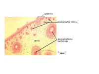
[[image: Section of Foetal Cat Skin.jpg|thumb|200px|right|A section of foetal cat skin showing developing hair follicles. ©RVC2008]] | [[image: Section of Foetal Cat Skin.jpg|thumb|200px|right|A section of foetal cat skin showing developing hair follicles. ©RVC2008]] | ||
| − | Hair germs begin from an aggregation of keratinocytes in the stratum basale of the [[Skin - Anatomy & Physiology#Epidermis|epidermis]]. The initiating factor is the underlying dermal fibroblast cells. The keratinocytes elongate, divide and relocate to the dermis. Dermal fibroblasts then | + | Hair germs begin from an aggregation of keratinocytes in the stratum basale of the [[Skin - Anatomy & Physiology#Epidermis|epidermis]]. The initiating factor is the underlying '''dermal fibroblast cells'''. The keratinocytes elongate, divide and relocate to the dermis. Dermal fibroblasts then form a '''dermal papilla''' beneath the hair germ. This causes stimulation of the basal stem cells to up-regulate their cycle, producing cells that will keratinise and form the hair shaft. Two swellings form on the shaft, one containing stem cells for follicle regeneration, the other becomes a [[Skin - Anatomy & Physiology#Glands|sebaceous gland]] which will secrete sebum onto the hair shaft. The follicles develop from an ectodermal bud which invades the mesenchyme during embryonic development. The mesoderm also condenses during the development creating an outer mesodermal component to the embedded part of the hair. |
==Structure and Function== | ==Structure and Function== | ||
| + | ===Hair growth cycle=== | ||
| + | Hair follicles grow in repeated cycles in a '''mosaic pattern''' so that the whole hair coat isn't lost at one time. Peaks of hair replacement occur in the '''spring and autumn'''. | ||
| + | |||
| + | *'''Anagen''': Growth phase. | ||
| + | The majority of hair follicles will be in this phase. The hair grows in length. | ||
| + | *'''Catagen''': Transition phase. | ||
| + | The dermal papilla is broken away and the follicle shrinks. | ||
| + | *'''Telogen''': Resting phase. | ||
| + | The hair doesn't grow but stays attached while the dermal papilla is resting. | ||
| + | |||
| + | After telogen the follicle re-enters anagen and the dermal papilla reattaches to the base of the follicle. If the old hair has not already epilated it will be pushed out by the new growing hair. | ||
| + | |||
| + | ===Control of hair growth=== | ||
| + | Hair growth is controlled by a number of factors including: '''photoperiod, ambient temperature, health, genetics, nutrition, hormones and local factors that directly influence the growth of hair follicles'''. | ||
| + | |||
| + | The most important factors controlling moulting are '''ambient temperature and photoperiod''', and this means that animals kept indoors in a near-constant environment may shed all year-round. | ||
| + | |||
| + | ===Hair and hair follicle anatomy=== | ||
| + | All hair has a similar structure: an outer cuticle, a cortex containing pigmentation, an inner medulla and a root or bulb from which the hair grows out of the follicle to form the shaft. | ||
| + | |||
| + | Each hair follicle has a sebaceous gland and an arrector pili muscle associated with it. | ||
| + | |||
| + | |||
| + | Hair follicles are divided into three parts: | ||
| + | :The ''infundibulum'' corresponds to the area from the opening of the sebaceous duct to the surface of the skin. | ||
| + | :The ''isthmus'' is the area between the opening of the sebaceous duct and the attachment of the arrector pili muscle. | ||
| + | :The ''inferior segment'' extends from the attachment of the arrector pili muscle to the dermal hair papilla. | ||
| + | |||
| + | |||
| + | The hair follicle originates from a peg of epidermal cells that grows down into the underlying dermis, where it forms a hair cone over a piece of dermis called the '''dermal papilla'''. The papilla provides the blood and nerve supply for the growing hair. From the hair cone, the cells keratinise and form a hair. As the hair grows up through the epidermis to the skin's surface, the cells at the point of the cone die, forming a channel, the hair follicle. | ||
| + | |||
===Hair Types=== | ===Hair Types=== | ||
| Line 11: | Line 43: | ||
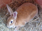
[[image: Rex rabbit.jpg|thumb|175px|left|The short, fine and densely packed guard hairs of a rex rabbit giving the coat a "velvet" appearance. ©Rachael Wallace2008]] | [[image: Rex rabbit.jpg|thumb|175px|left|The short, fine and densely packed guard hairs of a rex rabbit giving the coat a "velvet" appearance. ©Rachael Wallace2008]] | ||
| − | These are generally stiff and straight and form the outer haircoat of an animal. They are uniformly distributed across the skin surface and give the haircoat a smooth appearance. The smoothness of the coat is important in allowing rain to fall from the surface without penetrating deeper to the epidermis and causing loss of body temperature. The oily coating of the haircoat comes from the secretions of the sebaceous glands in the epidermis, associated with the hair follicle. This also contributes to the ‘waterproofing’ of the haircoat. Each hair consists of an outer cuticle, with a cortex and innermost medulla. It is composed of highly keratinised, dead epithelial cells, with the arrangement into the 3 layers conferring flexibility. | + | These are generally '''stiff and straight''' and form the outer haircoat of an animal. They are uniformly distributed across the skin surface and give the haircoat a '''smooth appearance'''. The smoothness of the coat is important in allowing '''rain to fall from the surface''' without penetrating deeper to the epidermis and causing loss of body temperature. The oily coating of the haircoat comes from the secretions of the sebaceous glands in the epidermis, associated with the hair follicle. This also contributes to the '''‘waterproofing’''' of the haircoat. Each hair consists of an outer cuticle, with a cortex and innermost medulla. It is composed of highly keratinised, dead epithelial cells, with the arrangement into the 3 layers conferring flexibility. |
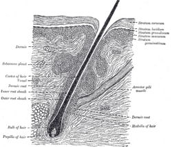
[[image: Section of hair follicle.jpg|thumb|250px|right|Section of skin showing a hair follicle. From Gray's Anatomy]] | [[image: Section of hair follicle.jpg|thumb|250px|right|Section of skin showing a hair follicle. From Gray's Anatomy]] | ||
| − | The '''''arrector pili''''' muscle is attached to the proximal end of the follicle and the dermis close to the point at which the hair is externalised. Involuntary contraction of this muscle in cold ambient temperatures causes erection of the hairs to trap warm air against the skin, thus providing [[Thermoregulation in Skin - Anatomy & Physiology|insulation]]. This can also be induced by the ‘fight or flight’ mechanism of the '''sympathetic nervous system''' | + | The '''''arrector pili''''' muscle is attached to the proximal end of the follicle and the dermis close to the point at which the hair is externalised. Involuntary contraction of this muscle in cold ambient temperatures causes '''erection of the hairs to trap warm air against the skin''', thus providing [[Thermoregulation in Skin - Anatomy & Physiology|insulation]]. This can also be induced by the ‘fight or flight’ mechanism of the '''sympathetic nervous system'''. |
| − | |||
| − | |||
*'''Wool hairs''' | *'''Wool hairs''' | ||
| − | These are thin, wavy, soft hairs that form the undercoat of animals. They are often much more numerous than the tough guard hairs and usually shorter. | + | These are thin, wavy, soft hairs that form the '''undercoat of animals'''. They are often much more numerous than the tough guard hairs and usually shorter. Their number increases in '''winter''' when they serve to keep the body warm. The thickness of the undercoat '''varies between species'''. Huskies have a very thick coat whereas Dobermans have almost no undercoat and cannot withstand cold weather very well. Wool hairs often '''share a single follicle with guard hairs''', although this is not common to all species. |
*'''Tactile hairs''' | *'''Tactile hairs''' | ||
| Line 26: | Line 56: | ||
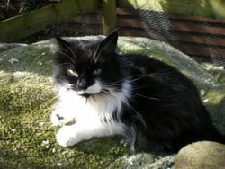
[[image: Long vibrissae.jpg|thumb|250px|left|Long vibrissae of a domestic cat. ©Rachael Wallace2008]] | [[image: Long vibrissae.jpg|thumb|250px|left|Long vibrissae of a domestic cat. ©Rachael Wallace2008]] | ||
| − | Tactile hairs are associated with a sensory function and are much thicker and stiffer than other hairs. They are mainly found on the face and can also be referred to as '''''whiskers''''' or '''''vibrissae'''''. Their follicles are much deeper into the | + | Tactile hairs are associated with a sensory function and are much '''thicker and stiffer''' than other hairs. They are mainly found on the face and can also be referred to as '''''whiskers''''' or '''''vibrissae'''''. Their '''follicles are much deeper''' into the hypodermis than guard or wool hairs and possess a '''venous sinus and nerve endings with mechanoreceptors'''. During embryonic development, they appear before other hair types. |
| + | These hairs are mostly found on the '''face, near the eyes and on the upper lip'''. However they are also found in other areas, for example in a cluster on the '''carpus in cats''', in a tuft on a '''dogs' cheeks'''. | ||
| + | |||
| + | *'''Tylotrich hairs''' | ||
| + | These large hair follicles are '''rapidly adapting mechanoreceptors'''. They may be present anywhere on the body. These structures consist of a single large hair, surrounded by neurovascular tissue at the level of the sebaceous gland. | ||
| + | |||
| + | {{Learning | ||
| + | |flashcards = [[Hair flashcards - Anatomy & Physiology|Hair Flashcards]]<br>[[Ear flashcards - Anatomy & Physiology|Ear Flashcards]]<br>[[Small Animal Dermatology Q&A 07]]<br>[[Small Animal Dermatology Q&A 03]] | ||
| + | |dragster = [[Integumentary System Histology Resource (III)|Hair Follicle Histology Dragster I]]<br>[[Integumentary System Histology Resource (IV)|Hair Follicle Histology Dragster II]]<br>[[Integumentary System Histology Resource (V)|Sinus Hair Histology Dragster I]]<br>[[Integumentary System Histology Resource (VI)|Sinus Hair Histology Dragster II]] | ||
| + | }} | ||
| − | == | + | ==References== |
| + | Aspinall, V. (2009) '''Introduction to veterinary anatomy and physiology''' ''Elsevier Health Sciences'' | ||
| − | + | Bowden, S. (2003) '''Veterinary anatomy and physiology: a workbook for students''' ''Elsevier Health Sciences'' | |
| − | + | Moriello, K (2005) '''Self-Assessment Colour Review - Small Animal Dermatology''' ''Manson'' | |
| + | ==Webinars== | ||
| + | <rss max="10" highlight="none">https://www.thewebinarvet.com/internal-medicine/webinars/feed</rss> | ||
[[Category:Integumentary System - Anatomy & Physiology]][[Category:Image Review]] | [[Category:Integumentary System - Anatomy & Physiology]][[Category:Image Review]] | ||
Latest revision as of 20:44, 3 January 2023
Development
Hair germs begin from an aggregation of keratinocytes in the stratum basale of the epidermis. The initiating factor is the underlying dermal fibroblast cells. The keratinocytes elongate, divide and relocate to the dermis. Dermal fibroblasts then form a dermal papilla beneath the hair germ. This causes stimulation of the basal stem cells to up-regulate their cycle, producing cells that will keratinise and form the hair shaft. Two swellings form on the shaft, one containing stem cells for follicle regeneration, the other becomes a sebaceous gland which will secrete sebum onto the hair shaft. The follicles develop from an ectodermal bud which invades the mesenchyme during embryonic development. The mesoderm also condenses during the development creating an outer mesodermal component to the embedded part of the hair.
Structure and Function
Hair growth cycle
Hair follicles grow in repeated cycles in a mosaic pattern so that the whole hair coat isn't lost at one time. Peaks of hair replacement occur in the spring and autumn.
- Anagen: Growth phase.
The majority of hair follicles will be in this phase. The hair grows in length.
- Catagen: Transition phase.
The dermal papilla is broken away and the follicle shrinks.
- Telogen: Resting phase.
The hair doesn't grow but stays attached while the dermal papilla is resting.
After telogen the follicle re-enters anagen and the dermal papilla reattaches to the base of the follicle. If the old hair has not already epilated it will be pushed out by the new growing hair.
Control of hair growth
Hair growth is controlled by a number of factors including: photoperiod, ambient temperature, health, genetics, nutrition, hormones and local factors that directly influence the growth of hair follicles.
The most important factors controlling moulting are ambient temperature and photoperiod, and this means that animals kept indoors in a near-constant environment may shed all year-round.
Hair and hair follicle anatomy
All hair has a similar structure: an outer cuticle, a cortex containing pigmentation, an inner medulla and a root or bulb from which the hair grows out of the follicle to form the shaft.
Each hair follicle has a sebaceous gland and an arrector pili muscle associated with it.
Hair follicles are divided into three parts:
- The infundibulum corresponds to the area from the opening of the sebaceous duct to the surface of the skin.
- The isthmus is the area between the opening of the sebaceous duct and the attachment of the arrector pili muscle.
- The inferior segment extends from the attachment of the arrector pili muscle to the dermal hair papilla.
The hair follicle originates from a peg of epidermal cells that grows down into the underlying dermis, where it forms a hair cone over a piece of dermis called the dermal papilla. The papilla provides the blood and nerve supply for the growing hair. From the hair cone, the cells keratinise and form a hair. As the hair grows up through the epidermis to the skin's surface, the cells at the point of the cone die, forming a channel, the hair follicle.
Hair Types
- Guard hairs
These are generally stiff and straight and form the outer haircoat of an animal. They are uniformly distributed across the skin surface and give the haircoat a smooth appearance. The smoothness of the coat is important in allowing rain to fall from the surface without penetrating deeper to the epidermis and causing loss of body temperature. The oily coating of the haircoat comes from the secretions of the sebaceous glands in the epidermis, associated with the hair follicle. This also contributes to the ‘waterproofing’ of the haircoat. Each hair consists of an outer cuticle, with a cortex and innermost medulla. It is composed of highly keratinised, dead epithelial cells, with the arrangement into the 3 layers conferring flexibility.
The arrector pili muscle is attached to the proximal end of the follicle and the dermis close to the point at which the hair is externalised. Involuntary contraction of this muscle in cold ambient temperatures causes erection of the hairs to trap warm air against the skin, thus providing insulation. This can also be induced by the ‘fight or flight’ mechanism of the sympathetic nervous system.
- Wool hairs
These are thin, wavy, soft hairs that form the undercoat of animals. They are often much more numerous than the tough guard hairs and usually shorter. Their number increases in winter when they serve to keep the body warm. The thickness of the undercoat varies between species. Huskies have a very thick coat whereas Dobermans have almost no undercoat and cannot withstand cold weather very well. Wool hairs often share a single follicle with guard hairs, although this is not common to all species.
- Tactile hairs
Tactile hairs are associated with a sensory function and are much thicker and stiffer than other hairs. They are mainly found on the face and can also be referred to as whiskers or vibrissae. Their follicles are much deeper into the hypodermis than guard or wool hairs and possess a venous sinus and nerve endings with mechanoreceptors. During embryonic development, they appear before other hair types. These hairs are mostly found on the face, near the eyes and on the upper lip. However they are also found in other areas, for example in a cluster on the carpus in cats, in a tuft on a dogs' cheeks.
- Tylotrich hairs
These large hair follicles are rapidly adapting mechanoreceptors. They may be present anywhere on the body. These structures consist of a single large hair, surrounded by neurovascular tissue at the level of the sebaceous gland.
| Hair - Anatomy & Physiology Learning Resources | |
|---|---|
 Test your knowledge using drag and drop boxes |
Hair Follicle Histology Dragster I Hair Follicle Histology Dragster II Sinus Hair Histology Dragster I Sinus Hair Histology Dragster II |
 Test your knowledge using flashcard type questions |
Hair Flashcards Ear Flashcards Small Animal Dermatology Q&A 07 Small Animal Dermatology Q&A 03 |
References
Aspinall, V. (2009) Introduction to veterinary anatomy and physiology Elsevier Health Sciences
Bowden, S. (2003) Veterinary anatomy and physiology: a workbook for students Elsevier Health Sciences
Moriello, K (2005) Self-Assessment Colour Review - Small Animal Dermatology Manson
Webinars
Failed to load RSS feed from https://www.thewebinarvet.com/internal-medicine/webinars/feed: Error parsing XML for RSS


