Difference between revisions of "Protozoa"
(Redirected page to Category:Protozoa) |
|||
| (19 intermediate revisions by 3 users not shown) | |||
| Line 1: | Line 1: | ||
| − | + | {{toplink | |
| + | |backcolour = | ||
| + | |linkpage =Infectious agents and parasites | ||
| + | |linktext =INFECTIOUS AGENTS AND PARASITES | ||
| + | |pagetype=Bugs | ||
| + | |sublink1=Parasites | ||
| + | |subtext1=PARASITES | ||
| + | }} | ||
| + | |||
| + | ==Introduction== | ||
| + | [[Image:Balanditium.jpg|thumb|right|150px|Balantidium coli - trofozoite and cyst - Wikimedia Commons]] | ||
| + | [[Image:Flagella.jpg|thumb|right|150px|Flagella of ''E.coli'' - Nicolle Rager Fuller, National Science Foundation]] | ||
| + | All protozoa are unicellular eukaryotic organisms which store their genetic information in chromosomes in a nuclear envelope. Protozoa are classified depending on their structure and life cycle. This reflects the similarities of the diseases which they cause. Protozoa belong to the Animal Kingdom (rather than the Plant Kingdom) as they obtain their energy through the intake of organic material. | ||
| + | |||
| + | Protozoa usually range from 10μm-50μm but can grow up to 1mm. Thus, they are usually observed and classified using a microscope. | ||
| + | |||
| + | Protozoa multiply sexually, asexually and can also use a combination of both, as seen in the coccidia class. Replication can be by binary or multiple fission. Different protozoa use different forms of motility, including flagella, cilia, pseudopodia and gliding. | ||
| + | |||
| + | Not all protozoa are harmful. For example, the [[The Rumen - Anatomy & Physiology|rumen]] of ruminants and the [[Caecum - Anatomy & Physiology|caecum]] and [[Colon - Anatomy & Physiology|colon]] of horses are full of symbiotic protozoa. | ||
| + | |||
| + | ==Structure and function== | ||
| + | |||
| + | *Motile | ||
| + | |||
| + | *Protozoa possess all the 'usual' organelles which are found in most animal cells | ||
| + | **Nucleus, endoplasmic reticulum, mitochondria, Golgi bodies and lysosomes | ||
| + | |||
| + | *Protozoa also possess other cellular structures, organelles and sub-cellular structures which enable an '''independent existence''' to be led | ||
| + | |||
| + | *Cilia | ||
| + | **Fine, short hairs covering the protozoal surface each arising from a basal body | ||
| + | **Hairs beat in unison to enable the protozoa to move | ||
| + | **Wafts food towards the '''cytostome''' (mouth opening) | ||
| + | **E.g. ''Balantidium'' | ||
| + | |||
| + | *Flagellum | ||
| + | **Contractile fibre arising from a basal body | ||
| + | **Contracts in a whip like motion to propel protozoa | ||
| + | **Attached to body of some protozoa by an '''undulating membrane''' | ||
| + | **During movement, the organism's shape is maintained by microtubules in the pellicle | ||
| + | **E.g. ''Trypanosoma'' | ||
| + | |||
| + | *Pseudopodia | ||
| + | **Extensions of the cellular cytoplasm | ||
| + | **Cytoplasm flows into the pseudopodia allowing movement of the protozoa | ||
| + | **Also acts in a phagocytic manner surrounding food particles and enclosing it in a vacuole | ||
| + | **E.g. ''Entamoeba'' | ||
| + | |||
| + | *Gliding | ||
| + | **No obvious means of locomotion | ||
| + | **E.g. ''Eimeria'' | ||
| + | |||
| + | ==Nutrition and digestion== | ||
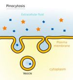
| + | [[Image:Pinocytosis.jpg|thumb|right|150px|Pinocytosis - Mariana Ruiz Villarreal]] | ||
| + | *Pinocytosis | ||
| + | **Droplets of fluid taken into the cell | ||
| + | **Generates small vesicles | ||
| + | **Usually used for extracellular fluid ingestion | ||
| + | **Requires ATP | ||
| + | |||
| + | *Phagocytosis | ||
| + | **Larger particles of matter taken into the cell | ||
| + | **Usually solid particles ingested | ||
| + | |||
| + | *Cell membrane envelops the fluid or food taking it into the cell | ||
| + | |||
| + | *Lysosomes fuse with the fluid/food vesicle initiating digestion | ||
| + | |||
| + | *Diffusion through the cell membrane allows excretion of metabolic products | ||
| + | |||
| + | ==Life Cycle== | ||
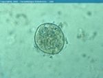
| + | [[Image:Balantidium pig trophozoite.jpg|thumb|right|150px|''Balantidium'' trophozoite from a pig - Joaquim Castellà Veterinary Parasitology Universitat Autònoma de Barcelona]] | ||
| + | |||
| + | *Most protozoal reproduction is asexual via binary fission, schizogony and sporogony | ||
| + | |||
| + | *Some protozoa also use sexual reproduction called gametogony | ||
| + | |||
| + | *In some species, sexual and asexual reproduction occurs in the same host, whilst in others asexual reproduction occurs in the vertebrate host and sexual reproduction in the arthropod vector | ||
| + | |||
| + | *Homoxenous | ||
| + | **Parasite uses a single host species during its life cycle (direct) | ||
| + | **E.g. ''Eimeria'' | ||
| + | |||
| + | *Heteroxenous | ||
| + | **Parasite uses more than one host during its life cycle (indirect) | ||
| + | **E.g. ''Trypanosomes'' | ||
| + | |||
| + | *Facultatively heteroxenous | ||
| + | **Parasite '''may''' use more than one host during its life cycle but this is not essential | ||
| + | **E.g. ''Toxoplasma gondii'' | ||
| + | |||
| + | ===Example of a Protozoal Life Cycle=== | ||
| + | [[Image:Coccidia.jpg|thumb|right|150px|Coccidia - Joel Mills]] | ||
| + | ''The following refers specifically to the life cycle of Coccidia spp.'' | ||
| + | *The '''oocyst''' is the resistant stage in the environment | ||
| + | |||
| + | *The infective '''sporozoite''' is released from the oocyst | ||
| + | |||
| + | *Inside the host, the sporozoites invade the intestinal epithelial tissue | ||
| + | **Sporozoites feed and grow | ||
| + | |||
| + | *As the sporozoite grows the nucleus divides forming a '''schizont''' | ||
| + | |||
| + | *The schizont contains numerous elongated '''merozoites''' | ||
| + | |||
| + | *The formation of merozoites is the first asexual reproductive stage called '''schizogony''' | ||
| + | |||
| + | *The schizont ruptures releasing the merozoites which also invade the epithelial cells | ||
| + | |||
| + | *Another generation of schizonts form which is the beginning of the sexual phase of reproduction called '''gametogony''' | ||
| + | |||
| + | *The merozoites form male '''microgamonts''' or female '''macrogamonts''' | ||
| + | **Collectively known as gamonts or gametocytes | ||
| + | |||
| + | *The microgamonts released from the microgametocyte penetrate and fertilise the macrogamont (which is contained within the macrogametocyte) | ||
| + | |||
| + | *Gametogony forms the '''zygote''' | ||
| + | **Surrounded by a cyst wall | ||
| + | **Forms the '''oocyst''' | ||
| + | |||
| + | *The oocyst is passed in the faeces and is unsporulated | ||
| + | |||
| + | *The oocyst becomes sporulated in the second asexual reproductive phase called '''sporogony''' | ||
| + | |||
| + | *Once the oocyst is sporulated it is infective | ||
| + | |||
| + | ==Protozoa of Veterinary Importance== | ||
| + | |||
| + | [[Coccidia]] | ||
| + | *''Eimeria'' | ||
| + | *''Isospora'' | ||
| + | |||
| + | [[Cryptosporidium|Cryptosporidium]] | ||
| + | |||
| + | [[Giardia]] | ||
| + | |||
| + | [[Piroplasmida]] | ||
| + | *Babesia | ||
| + | *Cytauxzoon | ||
| + | *Theileria | ||
| + | |||
| + | [[Tissue cyst-forming coccidia]] | ||
| + | *''Neospora'' | ||
| + | *''Sarcocystis'' | ||
| + | *''Toxoplasma'' | ||
| + | |||
| + | [[Tropical Protozoa]] | ||
| + | *''Leishmania'' spp. | ||
| + | *''Trypanosoma'' spp. | ||
| + | |||
| + | [[Other Important Protozoa]] | ||
| + | *''Balantidium'' | ||
| + | *''Cyclospora'' | ||
| + | *''Entamoeba'' | ||
| + | *''Histomonas meleagridis'' | ||
| + | *''Microsporidia'' | ||
| + | *''Tritrichomonas foetus'' | ||
| + | |||
| + | ==Useful Resources== | ||
| + | *http://www.veterinariavirtual.uab.es/parasito/diagnos003$/coproeq.htm | ||
| + | ''Brilliant microscopic pictures of protozoa and helminths'' | ||
| + | |||
| + | *http://www.vet.uga.edu/VPP/clerk/siegel/index.php | ||
| + | ''Detailed information and images, including clincial signs and pathogenesis, of East Coast Fever'' | ||
| + | |||
| + | *http://cal.vet.upenn.edu/projects/dxendopar/index.html#host | ||
| + | ''Useful online resource for diagnosing parasitic infections, courtesy of the Laboratory of Parasitology, University of Pennsylvania School of Veterinary Medicine'' | ||
Revision as of 09:31, 6 January 2009
|
|
Introduction
All protozoa are unicellular eukaryotic organisms which store their genetic information in chromosomes in a nuclear envelope. Protozoa are classified depending on their structure and life cycle. This reflects the similarities of the diseases which they cause. Protozoa belong to the Animal Kingdom (rather than the Plant Kingdom) as they obtain their energy through the intake of organic material.
Protozoa usually range from 10μm-50μm but can grow up to 1mm. Thus, they are usually observed and classified using a microscope.
Protozoa multiply sexually, asexually and can also use a combination of both, as seen in the coccidia class. Replication can be by binary or multiple fission. Different protozoa use different forms of motility, including flagella, cilia, pseudopodia and gliding.
Not all protozoa are harmful. For example, the rumen of ruminants and the caecum and colon of horses are full of symbiotic protozoa.
Structure and function
- Motile
- Protozoa possess all the 'usual' organelles which are found in most animal cells
- Nucleus, endoplasmic reticulum, mitochondria, Golgi bodies and lysosomes
- Protozoa also possess other cellular structures, organelles and sub-cellular structures which enable an independent existence to be led
- Cilia
- Fine, short hairs covering the protozoal surface each arising from a basal body
- Hairs beat in unison to enable the protozoa to move
- Wafts food towards the cytostome (mouth opening)
- E.g. Balantidium
- Flagellum
- Contractile fibre arising from a basal body
- Contracts in a whip like motion to propel protozoa
- Attached to body of some protozoa by an undulating membrane
- During movement, the organism's shape is maintained by microtubules in the pellicle
- E.g. Trypanosoma
- Pseudopodia
- Extensions of the cellular cytoplasm
- Cytoplasm flows into the pseudopodia allowing movement of the protozoa
- Also acts in a phagocytic manner surrounding food particles and enclosing it in a vacuole
- E.g. Entamoeba
- Gliding
- No obvious means of locomotion
- E.g. Eimeria
Nutrition and digestion
- Pinocytosis
- Droplets of fluid taken into the cell
- Generates small vesicles
- Usually used for extracellular fluid ingestion
- Requires ATP
- Phagocytosis
- Larger particles of matter taken into the cell
- Usually solid particles ingested
- Cell membrane envelops the fluid or food taking it into the cell
- Lysosomes fuse with the fluid/food vesicle initiating digestion
- Diffusion through the cell membrane allows excretion of metabolic products
Life Cycle
- Most protozoal reproduction is asexual via binary fission, schizogony and sporogony
- Some protozoa also use sexual reproduction called gametogony
- In some species, sexual and asexual reproduction occurs in the same host, whilst in others asexual reproduction occurs in the vertebrate host and sexual reproduction in the arthropod vector
- Homoxenous
- Parasite uses a single host species during its life cycle (direct)
- E.g. Eimeria
- Heteroxenous
- Parasite uses more than one host during its life cycle (indirect)
- E.g. Trypanosomes
- Facultatively heteroxenous
- Parasite may use more than one host during its life cycle but this is not essential
- E.g. Toxoplasma gondii
Example of a Protozoal Life Cycle
The following refers specifically to the life cycle of Coccidia spp.
- The oocyst is the resistant stage in the environment
- The infective sporozoite is released from the oocyst
- Inside the host, the sporozoites invade the intestinal epithelial tissue
- Sporozoites feed and grow
- As the sporozoite grows the nucleus divides forming a schizont
- The schizont contains numerous elongated merozoites
- The formation of merozoites is the first asexual reproductive stage called schizogony
- The schizont ruptures releasing the merozoites which also invade the epithelial cells
- Another generation of schizonts form which is the beginning of the sexual phase of reproduction called gametogony
- The merozoites form male microgamonts or female macrogamonts
- Collectively known as gamonts or gametocytes
- The microgamonts released from the microgametocyte penetrate and fertilise the macrogamont (which is contained within the macrogametocyte)
- Gametogony forms the zygote
- Surrounded by a cyst wall
- Forms the oocyst
- The oocyst is passed in the faeces and is unsporulated
- The oocyst becomes sporulated in the second asexual reproductive phase called sporogony
- Once the oocyst is sporulated it is infective
Protozoa of Veterinary Importance
- Eimeria
- Isospora
- Babesia
- Cytauxzoon
- Theileria
- Neospora
- Sarcocystis
- Toxoplasma
- Leishmania spp.
- Trypanosoma spp.
- Balantidium
- Cyclospora
- Entamoeba
- Histomonas meleagridis
- Microsporidia
- Tritrichomonas foetus
Useful Resources
Brilliant microscopic pictures of protozoa and helminths
Detailed information and images, including clincial signs and pathogenesis, of East Coast Fever
Useful online resource for diagnosing parasitic infections, courtesy of the Laboratory of Parasitology, University of Pennsylvania School of Veterinary Medicine


