|
|
| (18 intermediate revisions by 6 users not shown) |
| Line 1: |
Line 1: |
| | + | {{toplink |
| | + | |backcolour = f5fffa |
| | + | |linkpage =Yeast-like fungi |
| | + | |linktext =YEAST-LIKE FUNGI |
| | + | |sublink1 =Flash Cards - WikiBugs |
| | + | |subtext1 =WIKIBUGS FLASHCARDS |
| | + | |pagetype =Bugs |
| | + | }} |
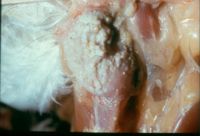
| | [[Image:Sour Crop.jpg|thumb|right|200px|Sour Crop - Copyright Professor Andrew N. Rycroft, BSc, PHD, C. Biol.F.I.Biol., FRCPath]] | | [[Image:Sour Crop.jpg|thumb|right|200px|Sour Crop - Copyright Professor Andrew N. Rycroft, BSc, PHD, C. Biol.F.I.Biol., FRCPath]] |
| − | ===Candidosis=== | + | ==<font color="purple">Candidosis</font>== |
| − | <FlashCard questions="4"> | + | {| border="3" cellpadding="8" |
| − | |q1=What are the three important species of ''Candida'' in animal infections?
| + | !width="400"|'''Question''' |
| − | |a1=
| + | !width="400"|'''Answer''' |
| − | *C. albicans
| + | !width="150"|'''Article''' |
| − | *C. tropicalis
| + | |- |
| − | *C. pelliculosa
| + | |<big>'''How do chromoblastomycosis infections spread in the host?''' |
| − | |l1=Candida_spp. | + | ||<font color="white"> <big> |
| − | |q2=What are the clinical signs of a sour crop infection?
| + | *'''''By the lymphatic system''''' |
| − | |a2=
| + | *'''''Disseminates to other tissues and organs''''' |
| − | *White-grey lesions in mouth, oesophagus and crop
| + | ||[[Yeast-like fungi#Candidosis|<span title="Answer article">Link to Answer Article</span>]] |
| − | *Thickened crop wall
| + | |} |
| − | *Yellow-white necrotic material in crop
| + | <br> |
| − | |l2=Candida_spp. | |
| − | |q3=What other conditions are ''Candida'' spp. implicated in?
| |
| − | |a3=
| |
| − | *Thrush in humans
| |
| − | *Metritis, pyometra and vaginitis in mares
| |
| − | *Genital candidiosis in stallions
| |
| − | *Bovine mastitis
| |
| − | *Porcine stomach ulcers
| |
| − | |l3=Candida_spp. | |
| − | |q4=How would you demonstrate a ''Candida'' infection?
| |
| − | |a4=
| |
| − | *Skin scrapings in 20% KOH for microscopy
| |
| − | *Lactophenol Cotton Blue and stained by Gram or Methylene Blue stain
| |
| − | *Grows on Blood agar and Sabouraud's Dextrose agar
| |
| − | *Gram positive, oval, thin-walled budding cells with hyphal fragments
| |
| − | |l4=Candida_spp.
| |
| − | </FlashCard>
| |
| − | | |
| − | ===Cryptococcosis===
| |
| − | <FlashCard questions="5">
| |
| − | |q1=What is the main pathogenic species of ''Cryptococcus''? | |
| − | |a1=C. neoformans | |
| − | |l1=Cryptococcosis | |
| − | |q2=Where is ''Cryptococcus'' found in the environment?
| |
| − | |a2=
| |
| − | *Pigeon droppings
| |
| − | *Fruit
| |
| − | *Milk
| |
| − | *Soil
| |
| − | |l2=Cryptococcosis
| |
| − | |q3=Which body systems are affected?
| |
| − | |a3= | |
| − | *Respiratory
| |
| − | *Visceral
| |
| − | *Ocular
| |
| − | *Skeletal
| |
| − | *Systemic
| |
| − | *Cutaneous
| |
| − | |l3=Cryptococcosis | |
| − | |q4=Where is the most common site of infection in cats?
| |
| − | |a4=The tip of the nose
| |
| − | |l4=Cryptococcosis
| |
| − | |q5=How would you demonstrate a cryptococcosis infection?
| |
| − | |a5=
| |
| − | *India Ink
| |
| − | *PAS
| |
| − | *Gram stain (positive)
| |
| − | *Grows on Blood agar and Sabouraud's Dextrose agar
| |
| − | *Carbohydrate assimilation tests
| |
| − | *Latex agglutination for antigen, complement fixation, ELISA and IFAT can be used
| |
| − | |l5=Cryptococcosis
| |
| − | </FlashCard> | |
| − | | |
| − | ===Geotrichosis===
| |
| − | <FlashCard questions="2">
| |
| − | |q1=Which organs are usually affected in geotrichosis infections?
| |
| − | |a1=
| |
| − | *Udder
| |
| − | *Mucous membranes
| |
| − | *Bronchi
| |
| − | *Lungs
| |
| − | |l1=Geotrichosis
| |
| − | |q2=True or False: Geotrichosis infections are usually diagnosed post-mortem
| |
| − | |a2=True
| |
| − | |l2=Geotrichosis
| |
| − | </FlashCard> | |
| − | | |
| − | ===Malassezia pachydermidis===
| |
| − | <FlashCard questions="2">
| |
| − | |q1=Where in the body is ''Malassezia pachydermidis'' usually found?
| |
| − | |a1=
| |
| − | *Oily areas | |
| − | *Ear canal
| |
| − | *Skin
| |
| − | |l1=Yeast-like fungi#Malassezia pachydermatis
| |
| − | |q2=What is the pathogenesis of ''Malassezia pachydermatis'' infections?
| |
| − | |a2=
| |
| − | *Malassezia spp are part of the normal skin flora in many animal species and overgrowth most commonly occurs secondary to a change in skin microclimate due to underlying disease processes like food allergy, atopic dermatitis, endocrinopathies or keratinization defects
| |
| − | Regional lesions on the muzzle, ears, interdigital area and the perianal region
| |
| − | *Infection can also be generalised
| |
| − | *Erythematous, hyperpigmented, lichenified and scaly lesions with alopecia
| |
| − | |l2=Yeast-like fungi#Malassezia pachydermidis | |
| − | </FlashCard>
| |
| − | | |
| − | ===Rhodotorula===
| |
| − | <FlashCard questions="1">
| |
| − | |q1=What infections of dogs and horses are ''Rhodotorula'' spp. involved in? | |
| − | |a1=
| |
| − | *Canine ear infections
| |
| − | *Equine uterine infections
| |
| − | |l1=Rhodotorula
| |
| − | </FlashCard> | |
| − | | |
| − | ===Torulopsis glabrata===
| |
| − | <FlashCard questions="1">
| |
| − | |q1=What infections are ''Torulopsis'' spp. involved in?
| |
| − | |a1=
| |
| − | *Pyelonephritis, pneumonia, septicaemia and meningitis in humans
| |
| − | *Mastitis and abortion in cattle
| |
| − | *Systemic infection of monkeys and dogs
| |
| − | |l1=Torulopsis glabrata
| |
| − | </FlashCard> | |
| − | | |
| − | ===Trichosporonosis===
| |
| − | <FlashCard questions="2">
| |
| − | |q1=What infection is ''T. capitum'' involved in?
| |
| − | |a1=Bovine mastitis | |
| − | |l1=Trichosporonosis
| |
| − | |q2=What infections are ''Trichosporonosis beigelii'' involved in?
| |
| − | |a2=
| |
| − | *Feline nasal granuloma
| |
| − | *Skin infections in horses and monkeys
| |
| − | *Bovine and ovine mastitis
| |
| − | *Feline bladder infections
| |
| − | |l2=Trichosporonosis
| |
| − | </FlashCard> | |
| − | | |
| − | | |
| − | [[Category:Yeast-like Fungi]][[Category:Fungi Flashcards]]
| |
