Difference between revisions of "Male urogenital ultrasound - Donkey"
Jump to navigation
Jump to search
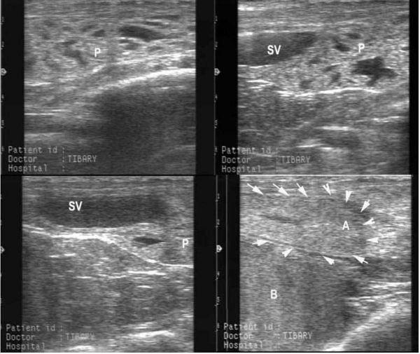
(New page: [[Image:Ultrasound jack genitals donkey.jpg|left|thumb|600px|<small><center>Ultrasonography of the internal genital organs in the jack: A) ampulla of the vas deferens, B) urinary bladder, ...) |
|||
| Line 4: | Line 4: | ||
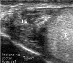
[[Image:Ultrasound testis donkey 2.jpg|right|thumb|250px|<small><center>Ultrasonography of the testis and epididymis. Caput (head) epididymis (HE) and testis (T). (Image courtesy of [http://drupal.thedonkeysanctuary.org.uk The Donkey Sanctuary])</center></small>]] | [[Image:Ultrasound testis donkey 2.jpg|right|thumb|250px|<small><center>Ultrasonography of the testis and epididymis. Caput (head) epididymis (HE) and testis (T). (Image courtesy of [http://drupal.thedonkeysanctuary.org.uk The Donkey Sanctuary])</center></small>]] | ||
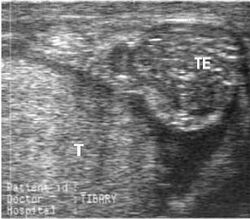
[[Image:Ultrasound testis donkey 3.jpg|right|thumb|250px|<small><center>Ultrasonography of the testis and epididymis. Cauda (tail) epididymis (TE) and testicular parenchyma (T). (Image courtesy of [http://drupal.thedonkeysanctuary.org.uk The Donkey Sanctuary])</center></small>]] | [[Image:Ultrasound testis donkey 3.jpg|right|thumb|250px|<small><center>Ultrasonography of the testis and epididymis. Cauda (tail) epididymis (TE) and testicular parenchyma (T). (Image courtesy of [http://drupal.thedonkeysanctuary.org.uk The Donkey Sanctuary])</center></small>]] | ||
| + | |||
| + | |||
| + | |||
| + | {{toplink | ||
| + | |titleborder=E0EEEE | ||
| + | |linkpage =Reproduction - Donkey | ||
| + | |linktext =Reproduction - Donkey | ||
| + | |sublink1 = Male Reproduction - Donkey | ||
| + | |subtext1 = Male Reproduction - Donkey | ||
| + | |pagetype = Donkey | ||
| + | }} | ||
| + | |||
| + | [[Category:Donkey]] | ||
| + | |||
| + | {{toplink | ||
| + | |titleborder=E0EEEE | ||
| + | |linkpage = Sponsors | ||
| + | |linktext = This page was sponsored and content provided by ''THE DONKEY SANCTUARY'' | ||
| + | }} | ||
Revision as of 22:40, 22 February 2010


|
|
|
|