Difference between revisions of "Adenoma"
Jump to navigation
Jump to search
Michuang0720 (talk | contribs) |
|||
| (13 intermediate revisions by 2 users not shown) | |||
| Line 1: | Line 1: | ||
| − | |||
| − | |||
[[Image:dogpap1.gif|right|thumb|100px|<small><center>Oral Papilloma Neoplasia in Dog (Courtesy of Alun Williams (RVC))</center></small>]] | [[Image:dogpap1.gif|right|thumb|100px|<small><center>Oral Papilloma Neoplasia in Dog (Courtesy of Alun Williams (RVC))</center></small>]] | ||
| − | + | *Adenomas are unusual but may develop in oropharyngeal salivary tissue. | |
| − | + | == Intestinal adenoma == | |
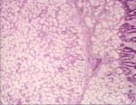
| − | + | [[Image:brunner gland adenoma.jpg|thumb|right|150px|Adenoma of brunners glands (duodenum) (Courtesy of Bristol BioMed Image Archive)]] | |
| − | [[Image: | ||
| − | |||
| − | |||
| − | |||
| − | + | * An adenoma is a growth of glandular origin. | |
| + | * Intestinal adenomas are found in both the [[Small Intestine - Anatomy & Physiology|small]] and [[Large Intestine - Anatomy & Physiology|large intestines]]. | ||
| + | * Intestinal adenomas usually grow into the lumen. | ||
| + | * These growths are bengin and polyp-like. | ||
| − | |||
| − | |||
| − | == | + | ==Tumours of the Perianal Area== |
| − | |||
| − | === | + | ===Hepatoid Gland Tumours (Perianal Adenomas)=== |
| − | + | [[Image:normal perianal gland.jpg|thumb|right|100px|Perianal gland- normal (Courtesy of Bristol BioMed Image Archive)]] * Affect the dog. | |
| + | * Arise from the solid, modified sebaceous circumanal glands. | ||
| + | * Common in ageing entire males. [[Image:perianal gland adenoma histopath.jpg|thumb|100px|Perianal gland- adenoma (Courtesy of Bristol BioMed Image Archive)]] | ||
| − | + | * Lesions range from hyperplasia to true adenomas (benign). | |
| − | + | ** These low grade lesions are under hormonal control. | |
| + | *** Castration/ administation of oestrogens or anti-androgens causes reduction in size.[[Image:perianal gland adenoma.jpg|thumb|right|100px|Perianal adenoma- gross appearance (Courtesy of Bristol BioMed Image Archive)]] | ||
| + | * Occasionally hepatoid carcinomas (malignant) arise in affected males | ||
| + | ** Outwith hormonal control. | ||
| + | * Hepatoid gland tumours occur rarely in bitches. | ||
| + | ** Are commonly malignant. | ||
| + | * Hepatoid glands are also found at the tail head, prepuce and occasionally other skin sites. | ||
| + | ** Hepatoid tumours can also arise in these areas. | ||
| − | + | ==Hepatocytic== | |
| + | *seen mostly in sheep and cattle | ||
| + | ===Gross=== | ||
| + | *a single, pale, soft, often large nodule | ||
| + | *well demarcated from adjacent tissue, often with a noticeable capsule | ||
| + | ===Microscopically=== | ||
| + | *normal hepatocytic appearance | ||
| + | *no portal tracts within the mass | ||
| + | *a capsule surrounds the growth | ||
| − | + | ==Cholangiocellular - bile duct== | |
| + | *very rare | ||
| + | *reported in dogs and cats | ||
| − | + | ==Pancreatic== | |
| − | |||
| − | |||
| − | |||
| − | |||
| − | |||
| − | |||
| − | |||
| − | |||
| − | |||
| − | |||
| − | |||
| − | |||
| − | |||
| − | |||
| − | |||
| − | |||
| − | |||
| − | |||
| − | |||
| − | |||
| − | |||
| − | |||
| − | |||
| − | |||
| − | |||
| − | |||
| − | |||
| − | |||
| − | |||
| − | |||
| − | |||
| − | |||
| − | |||
[http://w3.vet.cornell.edu/nst/nst.asp?Fun=Image&imgID=7754 Image of multifocal pancreatic adenoma in a dog from Cornell Veterinary Medicine] | [http://w3.vet.cornell.edu/nst/nst.asp?Fun=Image&imgID=7754 Image of multifocal pancreatic adenoma in a dog from Cornell Veterinary Medicine] | ||
| − | |||
| − | |||
| − | |||
| − | |||
| − | |||
| − | |||
| − | |||
| − | |||
| − | |||
| − | |||
| − | |||
| − | |||
| − | |||
| − | |||
| − | + | *Very rare | |
| + | *May be difficult to distinguish from nodular hyperplasia | ||
| + | *Single and larger nodules than normal [[Pancreas - Anatomy & Physiology|pancreas]] | ||
| − | |||
| − | |||
| − | |||
| − | |||
[[Category:Oropharynx - Pathology]] | [[Category:Oropharynx - Pathology]] | ||
[[Category:Intestines - Proliferative Pathology]] | [[Category:Intestines - Proliferative Pathology]] | ||
[[Category:Liver, Primary Tumours]] | [[Category:Liver, Primary Tumours]] | ||
[[Category:Pancreas_-_Hyperplastic_and_Neoplastic_Pathology]] | [[Category:Pancreas_-_Hyperplastic_and_Neoplastic_Pathology]] | ||
| + | [[Category:To_Do_-_Clinical]] | ||
[[Category:Neoplasia]] | [[Category:Neoplasia]] | ||
Revision as of 22:13, 28 June 2010
- Adenomas are unusual but may develop in oropharyngeal salivary tissue.
Intestinal adenoma
- An adenoma is a growth of glandular origin.
- Intestinal adenomas are found in both the small and large intestines.
- Intestinal adenomas usually grow into the lumen.
- These growths are bengin and polyp-like.
Tumours of the Perianal Area
Hepatoid Gland Tumours (Perianal Adenomas)
* Affect the dog.
- Arise from the solid, modified sebaceous circumanal glands.
- Common in ageing entire males.
- Lesions range from hyperplasia to true adenomas (benign).
- These low grade lesions are under hormonal control.
- Castration/ administation of oestrogens or anti-androgens causes reduction in size.
- These low grade lesions are under hormonal control.
- Occasionally hepatoid carcinomas (malignant) arise in affected males
- Outwith hormonal control.
- Hepatoid gland tumours occur rarely in bitches.
- Are commonly malignant.
- Hepatoid glands are also found at the tail head, prepuce and occasionally other skin sites.
- Hepatoid tumours can also arise in these areas.
Hepatocytic
- seen mostly in sheep and cattle
Gross
- a single, pale, soft, often large nodule
- well demarcated from adjacent tissue, often with a noticeable capsule
Microscopically
- normal hepatocytic appearance
- no portal tracts within the mass
- a capsule surrounds the growth
Cholangiocellular - bile duct
- very rare
- reported in dogs and cats
Pancreatic
Image of multifocal pancreatic adenoma in a dog from Cornell Veterinary Medicine
- Very rare
- May be difficult to distinguish from nodular hyperplasia
- Single and larger nodules than normal pancreas


