Difference between revisions of "Actinobacillosis - Cattle"
m (moved Actinobacillosis to Actinobacillosis - Cattle) |
|||
| (53 intermediate revisions by 2 users not shown) | |||
| Line 1: | Line 1: | ||
| − | + | {{unfinished}} | |
| − | + | {| cellpadding="10" cellspacing="0" border="1" | |
| + | | Also known as: | ||
| + | | '''Wooden tongue''' | ||
| + | |- | ||
| + | |} | ||
| − | |||
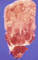
[[Image:Woodentongue1.gif|thumb|125px|<small><center>Fibrous Stroma - Cut Surface of actinobacillosis affected tongue (Courtesy of Alun Williams (RVC))</center></small>]] | [[Image:Woodentongue1.gif|thumb|125px|<small><center>Fibrous Stroma - Cut Surface of actinobacillosis affected tongue (Courtesy of Alun Williams (RVC))</center></small>]] | ||
| − | An infectious disease caused by the gram-negative coccobacilli | + | |
| + | ==Description== | ||
| + | An infectious disease caused by the gram-negative coccobacilli[[Actinobacillus lignieresii]]. Characterised by inflammation of the soft tissues of the head especially the [[Oral Cavity - Tongue - Anatomy & Physiology|tongue]] and [[Lymph Nodes - Anatomy & Physiology|pharangeal lymph nodes]] of cattle and sheep. The Causal agent is widespread in the environment and part of the normal flora of the GI mucosa. It gains acess to the tongue via small abrasions. It can be a progressive disease - low virulence but high persistence so the animal may stop eating and eventually die if not treated. | ||
| + | |||
==Signalment== | ==Signalment== | ||
| − | Occurs in cattle and sheep of all ages but particularly seen in young beef breeds | + | Occurs in cattle and sheep of all ages but particularly seen in young beef breeds particularly sucklers on poor forage. |
| + | |||
| + | ==Diagnosis== | ||
| + | A diagnosis can often be made on history and clinical signs. | ||
| + | Additionally, Pus from the abscesses may contain microcolonies interspersed with clublike pieces of calciuk phosphate which will give the appearance of sulphur granules. | ||
==Clinical Signs== | ==Clinical Signs== | ||
| − | Often begins like [[Foot and Mouth Disease|foot and mouth disease]]. Animals are dull, have difficulty | + | Often begins like [[Picornaviridae#Foot and Mouth Disease Virus|foot and mouth disease]]. Animals are dull, have difficulty masticating, inappetant and salivate profusely.. The tongue feels like a lump of wood especially the dorsal part of posterior 2/3rds which is often found in conjunction with small areas of ulceration on the side of [[Oral Cavity - Tongue - Anatomy & Physiology|tongue]]. |
| + | In contrast to foot and mouth cases are nearly always sporadic. | ||
| − | |||
| − | Occasionally generalised infections | + | Occasionally can see generalised infections but more commonly spreads to local lymph nodes of [[Alimentary - Anatomy & Physiology|alimentary tract]]and can affect soft tissue anywhere in the gastrointestinal tract including the rumen and reticulum. This may present as a chronic condition. |
| + | Additionally a cutaneous form has been reported where ulcers and nodules are present in the subcutaneous tissue containing yellow-green pus. | ||
| − | |||
| − | + | ====Pathogenesis==== | |
| − | + | *Very similar to [[Mandibular Osteomyelitis|Mandibular Osteomyelitis]] ("lumpy jaw"). | |
| − | |||
| − | |||
| − | |||
| − | + | ====Pathology==== | |
| + | [[Image:woodentongue2.gif|right|thumb|125px|<small><center>'Sulpher body' of Actinobacillosis (Courtesy of Alun Williams (RVC))</center></small>]] | ||
| − | |||
| − | |||
| − | |||
| − | + | *If cut into [[Oral Cavity - Tongue - Anatomy & Physiology|tongue]], substance of [[Oral Cavity - Tongue - Anatomy & Physiology|tongue]] changed to fibrous stroma with raised red nodules. (2-3mm across). | |
| + | *This lesion is a pyogenic granuloma with central '''sulphur body'''. | ||
| + | *Can spread from [[Oral Cavity - Tongue - Anatomy & Physiology|tongue]] to other tissues e.g. retropharyngeal [[Lymph Nodes - Anatomy & Physiology|lymph nodes]] and palate. | ||
| + | *This type of lesion is caused by the host response to the pathogen, rather than directly a pathogen effect. | ||
| − | |||
| − | |||
| − | |||
| − | |||
| − | |||
| − | |||
| − | |||
| − | |||
| − | + | *Small granulomatous lesions containing 'sulfa granules' of large numbers of gram-negative rods | |
| − | |||
| − | |||
| − | |||
| − | |||
| − | + | [[Category:Tongue_-_Pathology]][[Category:Cattle]] | |
| − | [[Category:Tongue_-_Pathology]][[Category: | + | [[Category:To Do - Caz]] |
| − | [[Category: | + | [[Category:Sheep]] |
| − | [[Category: | ||
| − | |||
Revision as of 12:46, 27 July 2010
| This article is still under construction. |
| Also known as: | Wooden tongue |
Description
An infectious disease caused by the gram-negative coccobacilliActinobacillus lignieresii. Characterised by inflammation of the soft tissues of the head especially the tongue and pharangeal lymph nodes of cattle and sheep. The Causal agent is widespread in the environment and part of the normal flora of the GI mucosa. It gains acess to the tongue via small abrasions. It can be a progressive disease - low virulence but high persistence so the animal may stop eating and eventually die if not treated.
Signalment
Occurs in cattle and sheep of all ages but particularly seen in young beef breeds particularly sucklers on poor forage.
Diagnosis
A diagnosis can often be made on history and clinical signs. Additionally, Pus from the abscesses may contain microcolonies interspersed with clublike pieces of calciuk phosphate which will give the appearance of sulphur granules.
Clinical Signs
Often begins like foot and mouth disease. Animals are dull, have difficulty masticating, inappetant and salivate profusely.. The tongue feels like a lump of wood especially the dorsal part of posterior 2/3rds which is often found in conjunction with small areas of ulceration on the side of tongue. In contrast to foot and mouth cases are nearly always sporadic.
Occasionally can see generalised infections but more commonly spreads to local lymph nodes of alimentary tractand can affect soft tissue anywhere in the gastrointestinal tract including the rumen and reticulum. This may present as a chronic condition.
Additionally a cutaneous form has been reported where ulcers and nodules are present in the subcutaneous tissue containing yellow-green pus.
Pathogenesis
- Very similar to Mandibular Osteomyelitis ("lumpy jaw").
Pathology
- If cut into tongue, substance of tongue changed to fibrous stroma with raised red nodules. (2-3mm across).
- This lesion is a pyogenic granuloma with central sulphur body.
- Can spread from tongue to other tissues e.g. retropharyngeal lymph nodes and palate.
- This type of lesion is caused by the host response to the pathogen, rather than directly a pathogen effect.
- Small granulomatous lesions containing 'sulfa granules' of large numbers of gram-negative rods

