Difference between revisions of "Intra-Oral Radiography Equipment - Small Animal"
Michuang0720 (talk | contribs) |
|||
| (4 intermediate revisions by 2 users not shown) | |||
| Line 1: | Line 1: | ||
| + | {{OpenPagesTop}} | ||
==Necessary Equipment== | ==Necessary Equipment== | ||
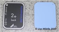
[[File:Digital plate.jpg|right|200px|thumb|Digital plate<small>''© Lisa Milella 2013''</small>]] | [[File:Digital plate.jpg|right|200px|thumb|Digital plate<small>''© Lisa Milella 2013''</small>]] | ||
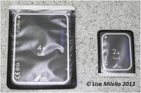
[[File:Digital phosphor plates.jpg|right|200px|thumb|Digital phosphor plates in a protective plastic cover. Occlusal and adult periapical sized films are shown.<small>''© Lisa Milella 2013''</small>]] | [[File:Digital phosphor plates.jpg|right|200px|thumb|Digital phosphor plates in a protective plastic cover. Occlusal and adult periapical sized films are shown.<small>''© Lisa Milella 2013''</small>]] | ||
| − | === | + | ===X-ray Generator=== |
| − | A '''standard veterinary | + | A '''standard veterinary x-ray machine''' can be used for taking dental radiographs but it has limitations. Often the x-ray machine is not in the same room where dental procedures are carried out and this means moving the patient. The tube head is mounted making positioning and adjustments in collimation and angulation difficult and the film-focus distance needs to be adjusted to between 30 and 50 cm which is not always possible with all machines.<br><br> |
| − | '''Dental | + | '''Dental x-ray generators''' have a mobile head and the collimation is confined by the cone. The cone also gives the film focal distance. The kV and mA are set, and the timer is the only adjustable control. Wall-mounted or mobile machines are available. |
<br><br> | <br><br> | ||
===Intra-Oral Film=== | ===Intra-Oral Film=== | ||
| − | Intra-oral or dental films should be used for taking dental radiographs. '''Intraoral films''' do not have | + | Intra-oral or dental films should be used for taking dental radiographs. '''Intraoral films''' do not have intensifying screens and thus need a '''high exposure'''. '''Dental film''' is available in '''different speeds'''. The sensitivity of the film determines the required exposure time – faster speed - higher sensitivity, requires less radiation to expose the film. Film sensitivity is increased by using larger silver halide crystals in the emulsion. As a result, E and F speed films produce images with a slightly lower image quality. The decreased image quality only becomes significant when viewed using magnification. The most commonly used speeds in veterinary dentistry are speed D (ultra) and speed E (ekta). Speed D is the equivalent of other non-screen film but speed E is rated at twice the speed of D, requiring half the exposure, with a small loss of quality. |
<br><br> | <br><br> | ||
Films are available in different sizes but the 3 most commonly used in dentistry are: | Films are available in different sizes but the 3 most commonly used in dentistry are: | ||
| Line 16: | Line 17: | ||
*Child periapical (2cm x 3.5cm) or size 1 | *Child periapical (2cm x 3.5cm) or size 1 | ||
<br><br> | <br><br> | ||
| − | Each film packet has individual wrapping to protect the film from light and moisture. The film inside the packet is wrapped in a black paper envelope. A lead foil covers the side of the film positioned away from the | + | Each film packet has individual wrapping to protect the film from light and moisture. The film inside the packet is wrapped in a black paper envelope. A lead foil covers the side of the film positioned away from the x-ray beam to protect against secondary radiation. |
<br><br> | <br><br> | ||
| − | Self contained films are also available in size 2 (periapical). The envelope contains the film with a separate compartment for developer and fixer. Films are relatively expensive but good when radiographs are only taken occasionally. The films unfortunately do not keep unless they are fixed for longer | + | Self contained films are also available in size 2 (periapical). The envelope contains the film with a separate compartment for developer and fixer. Films are relatively expensive but good when radiographs are only taken occasionally. The films unfortunately do not keep unless they are fixed for longer, otherwise they discolour with age. |
<br><br> | <br><br> | ||
Each film has a '''raised dot''' in one corner. The dot helps with orientation when viewing and mounting dental radiographs if the following procedure is adhered to. Firstly, the dot should face the incident beam. Secondly, the film should be placed in the [[Oral Cavity Overview - Anatomy & Physiology|mouth]] so that the dot is always facing a specific direction. We advise that dot is positioned so that it is always facing forward in the mouth.<br> | Each film has a '''raised dot''' in one corner. The dot helps with orientation when viewing and mounting dental radiographs if the following procedure is adhered to. Firstly, the dot should face the incident beam. Secondly, the film should be placed in the [[Oral Cavity Overview - Anatomy & Physiology|mouth]] so that the dot is always facing a specific direction. We advise that dot is positioned so that it is always facing forward in the mouth.<br> | ||
| − | Ensure the correct side of the film envelope is facing the incident beam. The envelope is marked or labelled. If exposed through the back of the envelope, the lead sheet will absorb much of the | + | Ensure the correct side of the film envelope is facing the incident beam. The envelope is marked or labelled. If exposed through the back of the envelope, the lead sheet will absorb much of the x-ray beam, resulting in an underexposed radiograph with the pattern of the lead sheet imposed on it. |
<br><br> | <br><br> | ||
==Processing== | ==Processing== | ||
| − | Films can be processed in a dark room using cups containing the processing chemicals or in a chair-side developer light-protected box. Some '''automatic processors''' will take dental film or separate automatic processors are available for dental film. The automatic processors deliver a fully fixed film, dried within 5 minutes. Ease of use and convenience are an advantage but cost and time to develop the films remains a disadvantage. A '''chair-side light box system''' is convenient to use and the films can be developed where you are working. Rapid processing chemicals are available resulting in a processed film within | + | Films can be processed in a dark room using cups containing the processing chemicals or in a chair-side developer light-protected box. Some '''automatic processors''' will take dental film or separate automatic processors are available for dental film. The automatic processors deliver a fully fixed film, dried within 5 minutes. Ease of use and convenience are an advantage but cost and time to develop the films remains a disadvantage. A '''chair-side light box system''' is convenient to use and the films can be developed where you are working. Rapid processing chemicals are available resulting in a processed film within one minute. The box is designed with a special transparent top that prevents light from entering and causing fogging of the film. Clips are available. Each film is opened, a clip attached to the corner then submerged in the developer for 15 seconds, washed, then fixed for 30 seconds then thoroughly washed under running water and hung up to dry. |
<br><br> | <br><br> | ||
| − | The films are too small to label individually but can be mounted in special mounts or stored in small envelopes with the client, patient and treatment details written | + | The films are too small to label individually but can be mounted in special mounts or stored in small envelopes with the client, patient and treatment details written on the envelope or mount. |
<br><br> | <br><br> | ||
==Digital Radiography== | ==Digital Radiography== | ||
Digital radiography refers to computer-generated images of radiographs. | Digital radiography refers to computer-generated images of radiographs. | ||
| − | '''Indirect methods''' of obtaining digital images make the exposure on a photostimulatable phosphor plate followed by | + | '''Indirect methods''' of obtaining digital images make the exposure on a photostimulatable phosphor plate followed by transfer of the latent image to a computer. |
'''Direct-to-digital equipment''' uses either a charge-coupled device (CCD) or a complementary metal oxide semiconductor (CMOS) sensor as a data input device that sends the x-ray image directly to a computer. This is the easiest and most time-efficient method. Most modern x-ray machines are compatible with most digital hardware and software packages. However, before purchasing either a new x-ray machine or a new digital sensor and software system, one should verify that the x-ray machine is capable of short enough exposure times for the digital equipment. Older x-ray machines cannot be used with digital sensors because most digital systems require exposure settings as low as 0.02 second for small patients. | '''Direct-to-digital equipment''' uses either a charge-coupled device (CCD) or a complementary metal oxide semiconductor (CMOS) sensor as a data input device that sends the x-ray image directly to a computer. This is the easiest and most time-efficient method. Most modern x-ray machines are compatible with most digital hardware and software packages. However, before purchasing either a new x-ray machine or a new digital sensor and software system, one should verify that the x-ray machine is capable of short enough exposure times for the digital equipment. Older x-ray machines cannot be used with digital sensors because most digital systems require exposure settings as low as 0.02 second for small patients. | ||
<br><br> | <br><br> | ||
Advantages of direct digital systems include the following: | Advantages of direct digital systems include the following: | ||
| − | * | + | *Immediate image availability |
| − | * | + | *Elimination of the use and disposal of developing and fixing fluids |
| − | * | + | *Elimination of errors during development |
*Allows digital enhancement to assist visualization | *Allows digital enhancement to assist visualization | ||
*Provides printed visit information for clients | *Provides printed visit information for clients | ||
| Line 47: | Line 48: | ||
| + | {{Lisa Milella written | ||
| + | |date = 1 October 2014}} | ||
| + | |||
| + | {{Learning | ||
| + | |Vetstream = [https://www.vetstream.com/felis/Content/Technique/teq60048.asp Dental radiography: overview] | ||
| + | }} | ||
| + | |||
| + | {{Waltham}} | ||
| + | |||
| + | {{OpenPages}} | ||
| + | |||
[[Category:Intra-Oral Radiography]] | [[Category:Intra-Oral Radiography]] | ||
| − | [[Category: | + | [[Category:Waltham reviewed]] |
Latest revision as of 19:11, 4 June 2016
Necessary Equipment
X-ray Generator
A standard veterinary x-ray machine can be used for taking dental radiographs but it has limitations. Often the x-ray machine is not in the same room where dental procedures are carried out and this means moving the patient. The tube head is mounted making positioning and adjustments in collimation and angulation difficult and the film-focus distance needs to be adjusted to between 30 and 50 cm which is not always possible with all machines.
Dental x-ray generators have a mobile head and the collimation is confined by the cone. The cone also gives the film focal distance. The kV and mA are set, and the timer is the only adjustable control. Wall-mounted or mobile machines are available.
Intra-Oral Film
Intra-oral or dental films should be used for taking dental radiographs. Intraoral films do not have intensifying screens and thus need a high exposure. Dental film is available in different speeds. The sensitivity of the film determines the required exposure time – faster speed - higher sensitivity, requires less radiation to expose the film. Film sensitivity is increased by using larger silver halide crystals in the emulsion. As a result, E and F speed films produce images with a slightly lower image quality. The decreased image quality only becomes significant when viewed using magnification. The most commonly used speeds in veterinary dentistry are speed D (ultra) and speed E (ekta). Speed D is the equivalent of other non-screen film but speed E is rated at twice the speed of D, requiring half the exposure, with a small loss of quality.
Films are available in different sizes but the 3 most commonly used in dentistry are:
- Occlusal film (5cm x 7cm) or size 4
- Adult periapical (3cm x 4cm) or size 2, and
- Child periapical (2cm x 3.5cm) or size 1
Each film packet has individual wrapping to protect the film from light and moisture. The film inside the packet is wrapped in a black paper envelope. A lead foil covers the side of the film positioned away from the x-ray beam to protect against secondary radiation.
Self contained films are also available in size 2 (periapical). The envelope contains the film with a separate compartment for developer and fixer. Films are relatively expensive but good when radiographs are only taken occasionally. The films unfortunately do not keep unless they are fixed for longer, otherwise they discolour with age.
Each film has a raised dot in one corner. The dot helps with orientation when viewing and mounting dental radiographs if the following procedure is adhered to. Firstly, the dot should face the incident beam. Secondly, the film should be placed in the mouth so that the dot is always facing a specific direction. We advise that dot is positioned so that it is always facing forward in the mouth.
Ensure the correct side of the film envelope is facing the incident beam. The envelope is marked or labelled. If exposed through the back of the envelope, the lead sheet will absorb much of the x-ray beam, resulting in an underexposed radiograph with the pattern of the lead sheet imposed on it.
Processing
Films can be processed in a dark room using cups containing the processing chemicals or in a chair-side developer light-protected box. Some automatic processors will take dental film or separate automatic processors are available for dental film. The automatic processors deliver a fully fixed film, dried within 5 minutes. Ease of use and convenience are an advantage but cost and time to develop the films remains a disadvantage. A chair-side light box system is convenient to use and the films can be developed where you are working. Rapid processing chemicals are available resulting in a processed film within one minute. The box is designed with a special transparent top that prevents light from entering and causing fogging of the film. Clips are available. Each film is opened, a clip attached to the corner then submerged in the developer for 15 seconds, washed, then fixed for 30 seconds then thoroughly washed under running water and hung up to dry.
The films are too small to label individually but can be mounted in special mounts or stored in small envelopes with the client, patient and treatment details written on the envelope or mount.
Digital Radiography
Digital radiography refers to computer-generated images of radiographs.
Indirect methods of obtaining digital images make the exposure on a photostimulatable phosphor plate followed by transfer of the latent image to a computer.
Direct-to-digital equipment uses either a charge-coupled device (CCD) or a complementary metal oxide semiconductor (CMOS) sensor as a data input device that sends the x-ray image directly to a computer. This is the easiest and most time-efficient method. Most modern x-ray machines are compatible with most digital hardware and software packages. However, before purchasing either a new x-ray machine or a new digital sensor and software system, one should verify that the x-ray machine is capable of short enough exposure times for the digital equipment. Older x-ray machines cannot be used with digital sensors because most digital systems require exposure settings as low as 0.02 second for small patients.
Advantages of direct digital systems include the following:
- Immediate image availability
- Elimination of the use and disposal of developing and fixing fluids
- Elimination of errors during development
- Allows digital enhancement to assist visualization
- Provides printed visit information for clients
- Automatically labels and stores images referenced to client and patient information
- Requires less radiation
- Templates for referral letters are integrated into most systems
| This article was written by Lisa Milella BVSc DipEVDC MRCVS. Date reviewed: 1 October 2014 |
| Intra-Oral Radiography Equipment - Small Animal Learning Resources | |
|---|---|
To reach the Vetstream content, please select |
Canis, Felis, Lapis or Equis |
| Endorsed by WALTHAM®, a leading authority in companion animal nutrition and wellbeing for over 50 years and the science institute for Mars Petcare. |
Error in widget FBRecommend: unable to write file /var/www/wikivet.net/extensions/Widgets/compiled_templates/wrt6934c7ed410e30_89269496 Error in widget google+: unable to write file /var/www/wikivet.net/extensions/Widgets/compiled_templates/wrt6934c7ed52d977_43535794 Error in widget TwitterTweet: unable to write file /var/www/wikivet.net/extensions/Widgets/compiled_templates/wrt6934c7ed5a6e44_85536132
|
| WikiVet® Introduction - Help WikiVet - Report a Problem |

