Difference between revisions of "Peripheral Odontogenic Fibroma"
(New page: {{unfinished}} {{dog}} ==Typical Signalment== ==Description== ==Diagnosis== ===Clinical Signs=== ===Diagnostic Imaging=== ===Biopsy=== ==Treatment== ==Prognosis== ==References...) |
|||
| (81 intermediate revisions by 5 users not shown) | |||
| Line 1: | Line 1: | ||
| − | {{ | + | {{OpenPagesTop}} |
| − | + | Also known as: ''''' Fibromatous epulis of periodontal ligament — Epulis — Ossifying Epulis | |
| − | + | ||
| + | ==Introduction== | ||
| + | [[File:Fibrous epulis.JPG|thumb|200px|right|Fibrous epulis]] | ||
| + | Peripheral odontogenic fibroma is a benign tumour that arises from the [[Tooth - Anatomy & Physiology#Periodontal Ligament|periodontal ligament]]. It was previously known as a fibromatous epulis and ossifying epulis depending on the degree of mineralization. | ||
| + | They present as firm, smooth swellings of the [[Gingiva|gingiva]] and are normally indistinguishable from [[Gingival Hyperplasia|gingival hyperplasia]]. | ||
==Typical Signalment== | ==Typical Signalment== | ||
| + | Most common benign tumour found in the oral cavity in dogs but is less common in cats. Is seen in dogs of any age but more common in those older than 6 years. . | ||
| − | == | + | ==Clinical Signs== |
| + | Include halitosis, oral bleeding, dental disruption or loss, facial or mandibular deformity, excessive salivation, growth protruding from the mouth and rarely dysphagia. | ||
| + | ==Diagnostic Imaging== | ||
| + | Radiographs are required to differentiate this benign neoplasm from malignant or locally aggressive lesions. Intra-oral radiographs will evaluate the oral lesion itself and thoracic radiography to evaluate for metastasies (if a malignancy is a diagnostic possibility). Radiographs typically show a soft tissue opacity in the the gingiva region with varying degrees of mineralization. Bone involvement is not a feature of this neoplasm and hence is not to be confused with [[Acanthomatous Ameloblastoma|Acanthomatous Ameloblastoma]] which often invades bone. | ||
| − | + | Radiography cannot be used to differentiate a peripheral odontogenic fibroma from a hyperplastic gingival lesion. | |
| − | == | + | ==Biopsy== |
| + | An incisional biopsy is required to obtain a definitive diagnosis. | ||
| − | == | + | ==Pathology== |
| − | + | [[Image:epulis.gif|right|thumb|200px|<small><center>Canine Epulis(Courtesy of Alun Williams (RVC))</center></small>]] | |
| − | + | Proliferation of fibrous tissue with a variety of osteoid, [[Tooth - Anatomy & Physiology#Cementum|cementum]] or [[Tooth - Anatomy & Physiology#Dentine|dentine]] like material. Isolated strands or islands of odontogenic epithelium are always present (ie: suggesting induction of connective tissue by the epithelial cells). | |
| + | The stroma contains neoplastic fibroblasts, with varying cellularity and the overlying epitheluim is normal. | ||
==Treatment== | ==Treatment== | ||
| + | A surgical excision of the neoplasm should be performed. The depth of the excision is determined by the location of the origin of the neoplasm at the periodontal ligament. | ||
| + | Excision may be at the gingival level or a deep resection involving the extraction of the affected tooth and curettage of the alveolar socket. | ||
| + | They do not recur if adequately excised. | ||
==Prognosis== | ==Prognosis== | ||
| + | Good following surgical resection. Recurrence is common following incomplete surgical resection. | ||
| + | |||
| + | {{Learning | ||
| + | |literature search = [http://www.cabdirect.org/search.html?q=title%3A%28%22Peripheral+Odontogenic+Fibroma%22%29+OR+%28title%3A%28epulis%29+AND+%28title%3A%28fibromatous%29+OR+title%3A%28ossifying%29%29%29 Peripheral Odontogenic Fibroma publications] | ||
| + | |Vetstream = [https://www.vetstream.com/felis/Content/Disease/dis60094.asp Odontoclastic tooth resorption] | ||
| + | }} | ||
==References== | ==References== | ||
| − | + | Tutt, C., Deeprose, J. and Crossley, D. (2007) '''BSAVA Manual of Canine and Feline Dentistry (3rd Edition)''' ''BSAVA'' | |
| + | |||
| + | Merck & Co (2008) '''The Merck Veterinary Manual (Eighth Edition)''' ''Merial'' | ||
| + | |||
| + | Verstraete, F.J.M., Ligthelmf, A.J. and Weber, A,(1992) '''The Histological Nature of Epulides in Dogs'''. Journal of comparative Pathology. (106) 169-182. | ||
| + | |||
| + | With thanks to Andrew Jefferies (Cambridge) and Alun Williams (RVC) for providing access to their lecture materials | ||
| + | |||
| + | |||
| + | {{review}} | ||
| + | |||
| + | {{OpenPages}} | ||
| − | + | [[Category:Teeth_-_Proliferative_Pathology]] | |
| + | [[Category:Neoplasia]] | ||
| + | [[Category:Expert_Review - Small Animal]] | ||
| + | [[Category:Oral Diseases - Dog]] | ||
| + | [[Category:Oral Diseases - Cat]] | ||
| + | [[Category:Bones - Pathology]] | ||
| + | [[Category:Oral Proliferations]] | ||
| + | [[Category:LisaM reviewing]] | ||
Latest revision as of 10:00, 21 May 2016
Also known as: Fibromatous epulis of periodontal ligament — Epulis — Ossifying Epulis
Introduction
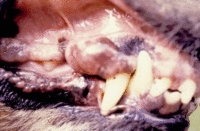
Peripheral odontogenic fibroma is a benign tumour that arises from the periodontal ligament. It was previously known as a fibromatous epulis and ossifying epulis depending on the degree of mineralization. They present as firm, smooth swellings of the gingiva and are normally indistinguishable from gingival hyperplasia.
Typical Signalment
Most common benign tumour found in the oral cavity in dogs but is less common in cats. Is seen in dogs of any age but more common in those older than 6 years. .
Clinical Signs
Include halitosis, oral bleeding, dental disruption or loss, facial or mandibular deformity, excessive salivation, growth protruding from the mouth and rarely dysphagia.
Diagnostic Imaging
Radiographs are required to differentiate this benign neoplasm from malignant or locally aggressive lesions. Intra-oral radiographs will evaluate the oral lesion itself and thoracic radiography to evaluate for metastasies (if a malignancy is a diagnostic possibility). Radiographs typically show a soft tissue opacity in the the gingiva region with varying degrees of mineralization. Bone involvement is not a feature of this neoplasm and hence is not to be confused with Acanthomatous Ameloblastoma which often invades bone.
Radiography cannot be used to differentiate a peripheral odontogenic fibroma from a hyperplastic gingival lesion.
Biopsy
An incisional biopsy is required to obtain a definitive diagnosis.
Pathology
Proliferation of fibrous tissue with a variety of osteoid, cementum or dentine like material. Isolated strands or islands of odontogenic epithelium are always present (ie: suggesting induction of connective tissue by the epithelial cells). The stroma contains neoplastic fibroblasts, with varying cellularity and the overlying epitheluim is normal.
Treatment
A surgical excision of the neoplasm should be performed. The depth of the excision is determined by the location of the origin of the neoplasm at the periodontal ligament. Excision may be at the gingival level or a deep resection involving the extraction of the affected tooth and curettage of the alveolar socket.
They do not recur if adequately excised.
Prognosis
Good following surgical resection. Recurrence is common following incomplete surgical resection.
| Peripheral Odontogenic Fibroma Learning Resources | |
|---|---|
To reach the Vetstream content, please select |
Canis, Felis, Lapis or Equis |
 Search for recent publications via CAB Abstract (CABI log in required) |
Peripheral Odontogenic Fibroma publications |
References
Tutt, C., Deeprose, J. and Crossley, D. (2007) BSAVA Manual of Canine and Feline Dentistry (3rd Edition) BSAVA
Merck & Co (2008) The Merck Veterinary Manual (Eighth Edition) Merial
Verstraete, F.J.M., Ligthelmf, A.J. and Weber, A,(1992) The Histological Nature of Epulides in Dogs. Journal of comparative Pathology. (106) 169-182.
With thanks to Andrew Jefferies (Cambridge) and Alun Williams (RVC) for providing access to their lecture materials
| This article has been peer reviewed but is awaiting expert review. If you would like to help with this, please see more information about expert reviewing. |
Error in widget FBRecommend: unable to write file /var/www/wikivet.net/extensions/Widgets/compiled_templates/wrt69a14eaa935515_76217366 Error in widget google+: unable to write file /var/www/wikivet.net/extensions/Widgets/compiled_templates/wrt69a14eaa9b9353_83088137 Error in widget TwitterTweet: unable to write file /var/www/wikivet.net/extensions/Widgets/compiled_templates/wrt69a14eaaa33bd3_10260602
|
| WikiVet® Introduction - Help WikiVet - Report a Problem |

