Difference between revisions of "Coccidiosis - Goat"
| (9 intermediate revisions by 3 users not shown) | |||
| Line 1: | Line 1: | ||
| − | [[Image:Goats.jpg|thumb|right|150px|Goats - nabrown RVC]] | + | {{OpenPagesTop}} |
| − | + | == Introduction == | |
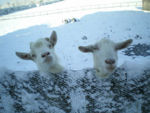
| + | [[Image:Goats.jpg|thumb|right|150px|Goats - nabrown RVC]] | ||
| + | Coccidiosis is the most common cause of diarrhoea in kids aged 3 weeks - 5 months of age and is particularly prevalent when housed. There are twelve known species of [[:Category:Coccidia|coccidia]] that affect goats and all but one are specific to goats with the other being transmissible between sheep and goats. All coccidia species affecting goats are of the [[Eimeria spp.|''Eimeria'' genus]]. | ||
| − | + | All goats are infected with coccidia in the first few weeks of life and in is thought that other factors such as hygiene, stress levels, transport and other concurrent infections lead to the disease becoming clinical. Goats then build up an immunity to the disease as they age, meaning they will excrete a small number of oocysts into the environment on a continuous basis. If the immune goat then becomes stressed then it is likely a relaxation in immunity will occur. | |
| − | |||
| − | * | + | |
| − | [[Category: | + | == Clinical Signs == |
| + | |||
| + | Watery or mucoid diarrhoea is usually present, which may be blood-tinged. Tenesmus may be present, but is not so common as in cattle or sheep. General signs of depression and malaise will also be seen in the affected animal, such as reluctance to eat, weight loss and weakness. If the infestation is particularly severe, sudden onset colic and collapse followed by death can occur in younger kids. | ||
| + | |||
| + | |||
| + | == Diagnosis == | ||
| + | |||
| + | History, clinical signs and signalment are useful in creating a differential list. Fresh faecal samples should be taken and observed microscopically for presence of oocysts. Oocysts should be counted, as all goats will excrete some oocysts but this does not mean coccidiosis is the cause of the disease. An oocyst count of 1,000,000 or more is usually indicative of clinical disease. | ||
| + | |||
| + | If an animal has died then a post mortem examination should be performed. Findings such as haemorrhagic enteritis in severe cases and small white focal lesions on the intestines may be noted. Smears should be taken from the mucosal surface of the damaged intestine to inspect for coccidia under the microscope. | ||
| + | |||
| + | |||
| + | == Treatment and Control == | ||
| + | |||
| + | A hygienic environment with good husbandry is the key to coccidia control. | ||
| + | |||
| + | *Avoid overcrowding | ||
| + | *Do not mix age groups | ||
| + | |||
| + | Other factors which will dramatically reduce clinical coccidiosis are avoiding faecal contamination of feed and water, deep cleaning of litter pen every month and clean bedding on a daily basis if possible. | ||
| + | |||
| + | If treatment is required in an outbreak, a drug must be used off licence as no anti-coccidials or coccidio-stats are licensed for use in goats. Sulphonamides, decoquinate, diclazuril and toltrazuril are commonly used to good effect. | ||
| + | |||
| + | Rehydration is usually required in a clinical case of coccidiosis and an electrolyte solution should be fed to the affected animals. Milk should be fed only in small amounts as the damage to the intestine means the milk will not be digested efficiently resulting in more diarrhoea. | ||
| + | |||
| + | |||
| + | == References == | ||
| + | |||
| + | Fox, M and Jacobs, D. (2007) Parasitology Study Guide Part 1: Ectoparasites Royal Veterinary College <br>Matthews, J. (2009) Disease of the Goat (Third Edition), Wiley Blackwell Publishing<br>Radostits, O.M, Arundel, J.H, and Gay, C.C. (2000) Veterinary Medicine: a textbook of the diseases of cattle, sheep, pigs, goats and horses Elsevier Health Sciences <br>Smith, M.C, Sherman, D.M. (2009) Goat Medicine (Second Edition) Wiley-Blackwell Publishing<br> | ||
| + | |||
| + | |||
| + | {{review}} | ||
| + | |||
| + | {{OpenPages}} | ||
| + | |||
| + | [[Category:Goat_Parasites]] [[Category:Alimentary Diseases - Goat]][[Category:Expert_Review - Farm Animal]] | ||
Latest revision as of 16:10, 30 July 2012
Introduction
Coccidiosis is the most common cause of diarrhoea in kids aged 3 weeks - 5 months of age and is particularly prevalent when housed. There are twelve known species of coccidia that affect goats and all but one are specific to goats with the other being transmissible between sheep and goats. All coccidia species affecting goats are of the Eimeria genus.
All goats are infected with coccidia in the first few weeks of life and in is thought that other factors such as hygiene, stress levels, transport and other concurrent infections lead to the disease becoming clinical. Goats then build up an immunity to the disease as they age, meaning they will excrete a small number of oocysts into the environment on a continuous basis. If the immune goat then becomes stressed then it is likely a relaxation in immunity will occur.
Clinical Signs
Watery or mucoid diarrhoea is usually present, which may be blood-tinged. Tenesmus may be present, but is not so common as in cattle or sheep. General signs of depression and malaise will also be seen in the affected animal, such as reluctance to eat, weight loss and weakness. If the infestation is particularly severe, sudden onset colic and collapse followed by death can occur in younger kids.
Diagnosis
History, clinical signs and signalment are useful in creating a differential list. Fresh faecal samples should be taken and observed microscopically for presence of oocysts. Oocysts should be counted, as all goats will excrete some oocysts but this does not mean coccidiosis is the cause of the disease. An oocyst count of 1,000,000 or more is usually indicative of clinical disease.
If an animal has died then a post mortem examination should be performed. Findings such as haemorrhagic enteritis in severe cases and small white focal lesions on the intestines may be noted. Smears should be taken from the mucosal surface of the damaged intestine to inspect for coccidia under the microscope.
Treatment and Control
A hygienic environment with good husbandry is the key to coccidia control.
- Avoid overcrowding
- Do not mix age groups
Other factors which will dramatically reduce clinical coccidiosis are avoiding faecal contamination of feed and water, deep cleaning of litter pen every month and clean bedding on a daily basis if possible.
If treatment is required in an outbreak, a drug must be used off licence as no anti-coccidials or coccidio-stats are licensed for use in goats. Sulphonamides, decoquinate, diclazuril and toltrazuril are commonly used to good effect.
Rehydration is usually required in a clinical case of coccidiosis and an electrolyte solution should be fed to the affected animals. Milk should be fed only in small amounts as the damage to the intestine means the milk will not be digested efficiently resulting in more diarrhoea.
References
Fox, M and Jacobs, D. (2007) Parasitology Study Guide Part 1: Ectoparasites Royal Veterinary College
Matthews, J. (2009) Disease of the Goat (Third Edition), Wiley Blackwell Publishing
Radostits, O.M, Arundel, J.H, and Gay, C.C. (2000) Veterinary Medicine: a textbook of the diseases of cattle, sheep, pigs, goats and horses Elsevier Health Sciences
Smith, M.C, Sherman, D.M. (2009) Goat Medicine (Second Edition) Wiley-Blackwell Publishing
| This article has been peer reviewed but is awaiting expert review. If you would like to help with this, please see more information about expert reviewing. |
Error in widget FBRecommend: unable to write file /var/www/wikivet.net/extensions/Widgets/compiled_templates/wrt693a1684df8548_04113825 Error in widget google+: unable to write file /var/www/wikivet.net/extensions/Widgets/compiled_templates/wrt693a1684efc506_52619606 Error in widget TwitterTweet: unable to write file /var/www/wikivet.net/extensions/Widgets/compiled_templates/wrt693a16850a3e88_08820678
|
| WikiVet® Introduction - Help WikiVet - Report a Problem |
