Difference between revisions of "Category:Stomach and Abomasum - Inflammatory Pathology"

From WikiVet English
Jump to navigation Jump to search
Line 1: Line 1:
 
 
* '''Gastritis''' refers to inflammation of the [[Forestomach - Anatomy & Physiology|stomach]].
 
* '''Gastritis''' refers to inflammation of the [[Forestomach - Anatomy & Physiology|stomach]].
  
Line 8: Line 7:
 
===[[Oedema Disease]] In The Pig===
 
===[[Oedema Disease]] In The Pig===
  
==Erosive and Ulcerative Gastritis ==
 
 
* Causes '''gastric ulcers'''
 
* Seen
 
** Commonly in the dog and pig.
 
** In young calves weaned onto a coarse diet.
 
** These usually heal as animal gets older.
 
** In the horse, associated with parasites.
 
* Once started, gastric ulcers can erode deeply.
 
** May penetrate gastric wall leading to peritonitis.
 
** May erode a blood vessel to cause haemorrhage.
 
 
===Pathology===
 
 
====Gross====
 
[[Image:gastric ulcer.jpg|thumb|right|150px|Gastic ulcer- gross (Courtesy of BioMed Image Archive)]]
 
* Round or oval lesions from 1-4 cm in diameter.
 
* Sharply “punched out” lesions with perpendicular or slightly overhanging walls.
 
* Borders are level with, or slightly raised above, the surrounding mucosa.
 
* Depth is variable.
 
** Some penetrate the superficial mucosa only.
 
** Some deeply penetrate the muscularis externa.
 
* Base may be markedly haemorrhagic.
 
** In advanced chronic cases, scarring may result in a puckered appearance.
 
 
====Histological====
 
[[Image:gastric ulcer histopath.jpg|thumb|right|150px|Gastric ulcer- histological (Courtesy of BioMed Image Archive)]]
 
* Appearance varies with the degree aggressiveness of the ulcer and the amount of healing which has occurred.
 
** Rapidly excavating ulcers have minimal granulation tissue and collagen deposition.
 
** Others may have a necrotic base with a framework of granulation tissue and collagen.
 
* The blood vessels at the base of the ulcer may be thickened and thrombosed.
 
* In the bovine, the ulcer may have a superimposed fungal infection.
 
 
===Pathogenesis===
 
 
* '''There are differences in pathogenesis between species.'''
 
 
====Cattle====
 
 
* Management-related in young calves and dairy cows.
 
* May also be caused by infectious agents, e.g. [[Bovine Viral Diarrhoea Virus|mucosal disease/ bovine viral diarrhoea virus]].
 
* Ulcers have a tendency to bleed and perforate.
 
 
====Horse====
 
 
* Affects the pars oesophagea (margo plicatus) in adults and foals.
 
* Due to '''parasites''' - ''Gasterophilus'' (Bots).
 
* Bots are not as common as they once were.
 
* Look like big pink maggots.
 
* Killed by Ivermectin. 
 
* ''Gasterophilus'' leave large ulcers in glandular regions of the [[Forestomach - Anatomy & Physiology|stomach]].
 
** Ulcers / erosions are quite deep.
 
* The parasites are believed to be non-pathogenic, but in large numbers they probably produce some discomfort and poor growth.
 
* Carcinoma can also produce ulceration in the [[Forestomach - Anatomy & Physiology|stomach]] of the horse as, in other species.
 
 
* In foals, the glandular area may sometimes be affected.
 
** This may be e.g. stress-related, or due to used of NSAIDs.
 
 
====Dog====
 
 
* Although ulcers are often secondary to other diseases, primary idiopathic peptic ulcers do occur, due to
 
** Hyperacidity
 
** Gastric carcinoma in older dog


* Secondary ulcers are often associated with systemic diseases particularly '''uraemia''' and '''mast cell tumours'''. Gastric ulcer may be the cause of death but is not the primary disease. 
 
*# '''Mast cell tumours'''
 
*#*Boxers and Labradors are predisposed to these.
 
*#* Vomit continually together with abdominal pain.
 
*#* Ulcers are usually near the duodenum.
 
*#** Frequently secondarily infected.
 
*#** Often penetrate deeply.
 
*#* Actively secreting mast cell tumours produce histame, leasing to gastric hyperacidity and therefore secondary peptic ulcers.
 
*# '''Uraemia'''
 
*#* Gastric lesions usually occur with chronic renal disease.
 
*#** Gastrin is produced by the G cells of the gastric antrum during the gastric phase of digestion .
 
*#*** Acts on H2 receptors on parietal cells to increase production of HCl.
 
*#*** Increases release of histamine from gastric mucosal mast cells to increase HCl release.
 
*#** Serum levels of gastrin are increased in chronic renal disease in dogs and cats.
 
*#* In acute renal failure death ensues before gastric ulceration develops.
 
*#* '''Pathogenesis'''
 
*#** Loss of nephron and medullary concentration gradient in chronic interstitial nephritis mean collecting ducts cannot resorb fluid.
 
*#*** A common cause of interstitial nephritis in the dog was leptospirosis.
 
*#** Consequently, the animal drinks and urinates in enormous quantities, and urea is washed out with large quantities of fluid ("compensated renal failure").
 
*#** If fluid is restricted,  urea cannot be washed out and the animal becomes uraemic.
 
*#*** Urea is excreted into [[Forestomach - Anatomy & Physiology|stomach]], giving it a horrible ammoniacal smell and filling it with brown smelly liquid.
 
*#*** Urea is also excreted into the [[Colon - Anatomy & Physiology|colon]].
 
*#** Urea in the stomach breaks down to ammonia, irritating the mucosa and contributing to gastric ulcer.
 
*#** Uraemia also causes arteriolar degeneration in the submucosa, leading to hypoxic damage to the mucosa. This is another contributing factor to gastric ulcer.
 
*#** [[Control of Feeding - Anatomy & Physiology#The Vomit Reflex|Vomiting]] causes dehydration and further raises blood urea.
 
*#*** A vicious circle is produced-  ends in death by [[Control of Feeding - Anatomy & Physiology#The Vomit Reflex|vomiting]], dehydration and shock.
 
*#** '''Note:''' If an animal in compensated renal failure is given anaesthetic, it will not drink much. It then may start to [[Control of Feeding - Anatomy & Physiology#The Vomit Reflex|vomit]] and die due to uraemia.


* NSAIDs, Zollinger-Ellison syndrome (due to pancreatic gastrin-secreting tumour), cirrhosis and bile reflux can all also cause gastric ulcers in the dog.
 
 
====Pig====
 
 
* Gastic ulceration is quite common in the pig- May be seen in 50-60% of pigs arriving at slaughterhouses.
 
* Has serious economic consequences.
 
 
*'''Clinical'''
 
** Occasionally a well-grown pig will drop dead.
 
*** Deep ulcers have eroded into a blood vessel, causing massive haemorrhage into the [[Forestomach - Anatomy & Physiology|stomach]] from and producing death very rapidly.
 
** If long standing ulcers do not result in death, they do produce pain and discomfort.
 
*** Give low growth rate and poor feed conversion.
 
  
*'''Pathogenesis'''
+
[[:Category:Gastric Ulceration]]
** Gastric ulceration is associated with modern pig rearing, but the exact cause is unknown.
 
** Causes are associated with gastric hyperacidity, and gastric ulceration is probably a multifactorial disease.
 
** The following are suggested as possible causes:
 
*** Infection, e.g. ''Candida albicans'', ''Streptococci'', ''Staphylococci'' and mixes of these.
 
*** Copper toxicity- this is probably more significant.
 
**** Pigs are fed copper as growth promoter; 50 ppm is know to be toxic, and animals are often fed 250 ppm.
 
*** Vitamin E / Selenium deficiency.
 
*** Feeding on concrete floors.
 
**** Sand is licked up whe pigs eat.
 
*** Feeding finely milled cereal.
 
*** Stress
 
*** Possibly genetic factors.
 
  
*'''Pathology'''
 
** Most commonly affects pars oesophagea (squamous or non-glandular portion).
 
** Starts with hyperkeratosis in the stratum corneum
 
*** Appears rough and thickened
 
*** May stop at this stage.
 
** In approximately 30% of animals, the lesion starts to erode and quite deep ulcers may develop.
 
** In a significant small number ,very deep ulcers develop and may affect virtually all of pars oesophagea.
 
** Histologically, ulcers are large and flask-shaped ulcer with fibrin, necrosis, erosion and fibrosis at base.


==Fibrinous/ Diptheric Gastritis==
 
==Fibrinous/ Diptheric Gastritis==

Revision as of 12:58, 29 May 2010

  • Gastritis refers to inflammation of the stomach.


Gastritis, Catarrhal

Oedema Disease In The Pig

Category:Gastric Ulceration


Fibrinous/ Diptheric Gastritis

  • Not very common, but has severe consequences.
  • Dirty-white, crumbly fibrin is seen on the surface of mucosa.
  • Causes
    • Toxic
      • From drinking battery acid or other caustic material.
        • Also gives with stomatitis and oesophagitis as well.
      • Poisons such as mercuric chloride and carbolic acid also cause fibrinous/ diptheric gastritis.
    • Severe systemic disease
      • e.g. septicaemic Erysipelas and Swine Fever in pigs, or septicaemic Salmonellosis.
      • Not usually a primary problem but part of more severe generalised disease problem.


Haemorrhagic Gastritis

Clinical

  • Usually only seen post mortem.
  • Stomach full of thick tarry clots.
  • Occasionally will vomit blood in life.

Pathology

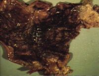
Haemorrhagic gastritis (Courtesy of BioMed Image Archive)

Gross

  • Wall of stomach is blacked and ulcerated.
    • Red, thickened, necrotic, haemorrhagic mucosa.

Histologically

  • Coagulative necrosis with fibrin, oedema, haemorrhage, and sometimes emphysema.
  • May extend deep into submucosa/muscle.

Pathogenesis

  • There are several causes of haemorrhagic gastritis
    1. Aspirin and non-steroidal anti inflammatory drug toxicity.
    2. Peracute / acute infections, e.g.
      • Swine Fever
      • Anthrax
      • Leptospirosis in dogs (Leptospira icterohaemorrhagiae).
    3. Clostridial disease
      • e.g. Braxy (Clostridium septicum)
        • Affects older lambs or yearlings producing sudden death.
        • Usually seen on sheep grazing on frosted grass so more common in colder areas.
        • Bacterial exotoxin causes acute abomasitis.
        • Pathology- At post mortem the stomach is grossly distended with partially clotted blood. The wall of the stomach is thickened,reddened and oedematous.
        • Diagnosed by isolation of organism from the stomach wall.
        • Is now usually vaccinated against (Heptovac 7 in 1 clostridial vaccine).
    4. Warfarin poisoning.

Vesicular Gastritis

  • Is not seen, as the stomach has no stratum spinosum.


Chronic gastritis

  • Chronic gastritis is usually proliferative rather any other type of gastric inflammation.
  • Usually a parasitic cause.
  • Occurs mostly in the pig and in cattle.
  • Pig
    • Redworms (Hyostrongylus)
    • Seen mostly in sows, and are present in up to 30% of pig herds.
    • Small numbers produce little pathology, but large numbers cause thin sow syndrome.
      • Animals eat well but slowly lose condition.
  • Cattle

Chronic Hypertrophic Gastritis In The Dog

  • Clinically see anorexia, weight loss, anaemia and associated hepatic disease.
  • Associated with protein loss into gut.

Pathology

  • Hyperplasia of mucosa.
    • Mucosa thrown up into folds.
    • Reduced numbers of parietal cells and increased numbers of goblet cells.

Chronic Atrophic Gastritis In The Dog

  • Aetiology uncertain.
  • Grossly: (may be difficult to appreciate)
    • Reduced mucosal thickness.
    • Loss of rugae.
  • Histologically
    • Mucosal thinning.
    • Loss of gastric glands.
    • Diffuse inflammatory infiltrate of lymphocytes and plasma cells.
    • Fewer eosinophils in lamina propria.

Subcategories

This category has only the following subcategory.

G