Difference between revisions of "Mucous Salivary Gland - Anatomy & Physiology"
Jump to navigation
Jump to search
m (Text replace - "Category:To Do - Review" to "Category:To Do - AP Review") |
|||
| Line 7: | Line 7: | ||
[[Category:Salivary Glands - Anatomy & Physiology]] | [[Category:Salivary Glands - Anatomy & Physiology]] | ||
[[Category:Oral Cavity and Oesophagus - Histology]] | [[Category:Oral Cavity and Oesophagus - Histology]] | ||
| − | [[Category:To Do - AimeeHicks]][[Category:To Do - Review]] | + | [[Category:To Do - AimeeHicks]][[Category:To Do - AP Review]] |
Revision as of 12:16, 18 October 2010
Overview
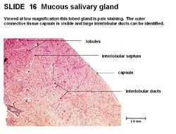
The mucous salivary gland has pale staining and is lobed. It has large, interlobular ducts in a connective tissue septum. It has an outer connective tissue capsule. The mucous acini produce a mucous secretion which is a viscous mix of glycoproteins. The cuboidal cells are filled with mucous droplets giving a ‘foamy’ appearance. The nucleus is displaced and flattened near the base of the cell. The mucous cells only stain faintly.
