Difference between revisions of "Sublingual Gland - Anatomy & Physiology"
Jump to navigation
Jump to search
m (Text replace - '|maplink = Alimentary (Concept Map)- Anatomy & Physiology' to '|maplink = ') |
|||
| Line 3: | Line 3: | ||
|linkpage =Alimentary - Anatomy & Physiology | |linkpage =Alimentary - Anatomy & Physiology | ||
|linktext =Alimentary System | |linktext =Alimentary System | ||
| − | |maplink = | + | |maplink = |
|pagetype =Anatomy | |pagetype =Anatomy | ||
|sublink1=Oral Cavity - Salivary Glands - Anatomy & Physiology#Major Salivary Glands | |sublink1=Oral Cavity - Salivary Glands - Anatomy & Physiology#Major Salivary Glands | ||
Revision as of 00:24, 3 July 2010
|
|
Sublingual Salivary Gland
- Merocrine secretion
- Smaller than parotid gland
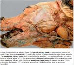
- Consists of monostomatic compact part drained by a single duct. Located over rostral mandibluar gland in dog. Duct runs with mandibluar gland duct and opens at the sublingual caruncle.
- Consists of polystomatic diffuse part drained by numerous smaller ducts. Thin strip below mucosa of the floor of oral cavity and duct opens beside the frenulum.
Histology
- Tubulo-acinar gland
- Mucous cells (stain lighter)
- Serous demilunes (stain darker)
- Demilunes secrete into lumen by canaliculi between mucous cells
Species Differences
Ruminant and Pig
Equine
- Diffuse part is the only part of the sublingual salivary gland present
