Mites
| This article has been peer reviewed but is awaiting expert review. If you would like to help with this, please see more information about expert reviewing. |
Astigmata (mites) introduction
Mites are one of the most successful and diverse vertebrate groups. The species of veterinary importance are parasitic, although a few non-parasitic mites are also important, such as oribatid mites (intermediate hosts for Anoplocephala and Moniezia) and forage mites.
Mites are very small, on average under 0.3mm in length and for this reason often go unnoticed. They spend the majority of their life cycle on the host and cause mange.
The body shows no segmentation but can be divided into two sections, the idiosoma and the gnathosoma. Adult and nymphs have four pairs of legs, whereas larvae have only three pairs of legs.
The taxonomy of mites is complex as there are over 8 families. In this article the families are split according to their location on the host into sub-surface (burrowing) and surface (non-burrowing) mites.
Burrowing Mites
- Sarcoptidae
Demodex
- Demodex spp. found on all domestic mammals and in humans
- Each host has its own species
- Causes demodecosis
Recognition
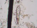
- Cigar shaped
- Four pairs of stumpy legs on the anterior end
- Long and narrow to fit into hair follicles
Life cycle
- Live as commensal organisms
- Live in hair follicles and in sebaceous glands
- Life cycle takes 3 weeks
Pathogenesis and epidemiology
Dogs
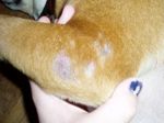
- Initial infection is slight hair loss which may resolve spontaneously or could spread over the body
- Squamous demodecosis
- Less serious
- Dry reaction
- Alopecia, desquamation and skin thickening
- Absent to mild pruritus
- Follicular/pustular demodecosis
- More serious
- Skin invasion by staphylococci
- Skin becomes wrinkled, thickened and contains pustules which ooze serum, blood or pus
- Affected animals may be seriously disfigured
- Severe pruritus is associated with secondary infection
- Immune factors are important in determining the severity and occurrence of demodecosis
- Familial susceptibility
- Immunosuppression
- Immunosuppressant therapy
Cats
- Rare
- Confined to the periocular region
- Mild squamous type only
Cattle
- Pea-sized nodules in the skin
- Each nodule contains several thousand mites
- Affects hide quality
- Economically important in Australia
Goats
- Becoming more common in goats
- Disease similar to that in cattle
Pigs, Sheep and Horses
- Rare
Diagnosis
- Liquid paraffin applied to a skin fold
- Deep skin scraping
Control
- Not easily accessible to acaricides due to their deep location in the skin
- Repeat treatments needed
- Recovery may take several months
- To aid acaricide penetration, clipping a dog's coat and washing is recommended
Notoedres
- Known as feline scabies
- Also common ectoparasite of tropical bats
- Parasite of cats, rats, man and rabbits
Recognition
- Similar to Sarcoptes
- Less distinct angles on body surface
- Females have suckers on legs 1 and 2
- Females are about 225μm in length and males 150μm
- Anal opening is distinctly dorsal (not posterior)
Pathogenesis
- Infection begins on the ear tips and spreads over the body
- Notoedres cati in notoedric skin infestation
- Causes dermatitis
- Burrowing of females damages keratinocytes leading to cytokine release
- Hypersensitivity reaction may occur
Diagnosis
- Superficial skin scraping
- A single nest in a scraping may yield many mites
Non-Burrowing Mites
- Live on the skin surface
- Feed on either skin scales and tissue or suck blood
Psoroptes
- Causes psoroptic skin infestation
Recognition
- Oval shaped
- Long legs
- Funnel shaped suckers on segmented pedicels
- 1-2mm in length
Life cycle
- Confined to skin surface
- Feed on serous exudate by siphoning
- Adult female can lay up to 100 eggs during her life time (1 month)
- 10 day life cycle
- 2 nymphal stages
Psoroptes cuniculi
- Parasite of rabbits
- Common among conventional rabbits
- Transmitted via contact
- Adapted to living in an aural environment
Pathogenesis
- The ears are painful and intensely pruritic
- Affected rabbits shake their heads and scratch their ears
- The inner surfaces of the pinnae are covered with brown, scaly, fetid material, and the skin beneath is raw
- Mites are grossly visible
- Histologically, there is chronic erosive and proliferative eosinophilic dermatitis
- The mites are non-burrowing and thus are found only in the exudate, not in the tissue
Diagnosis
- Microscopic examination for mites (low magnification)
- Appearance
Control
- Infestations are difficult to eliminate from a colony
- Ivermectin is usually effective
Psoroptes ovis
- Adult females are large mites at 750μm in length
- Males identified by copulatory suckers and paired posterior lobes
- Males attach to deutonymphs (second moult after larval stage) in a process called copula
- Males remain in copula until females moult for the last time
- Copulation occurs
- Life cycle last 14 days
- Transmitted by direct contact between sheep
- Indirect transmission can also occur
Pathogenesis
- Economically important ectoparasite of sheep
- Causes sheep scab
- Wool loss, restlessness, biting, scratching of infested area and decreased productivity through decreased weight gain
- Usually seen in late autumn and early winter (although may also occur in late summer)
- Population numbers decline after shearing due to a change in the micro-climate, then build up again as the fleece grows
- Notifiable in UK
- Mites found under scabs and in skin folds
- Lesions most common on flanks, neck, back and shoulders
- Causes pruritic condition of cattle
- Active in keratin layer
- Mouthparts abrade the skin
- Antigenic material in mite faeces can lead to hypersensitivity reactions
Diagnosis
- Skin scraping
- KOH added
- Warm slide over a bunsen flame
- Examine under a microscope
Treatment
- Sheep
- Plunge dipping; no less than 1 minute and must dip head at lease once
- Can treat with avermectins or milbemycins by injection
- Cattle, horses and rabbits
- No licensed product for horses in the UK
- Cattle and rabbits can be treated with avermectins, milbemycins or topical acaricides
Chorioptes bovis
- These cause parasitic skin infestation
- Surface parasite of horses and cattle
- Less pathogenic than Psoroptes
- Mouthparts cannot pierce the skin
- Life cycle takes 3 weeks
Recognition
- Oval body
- Long legs
- Cup shaped suckers on unsegmented pedicels
- Females about 300μm in length
Pathogenesis
- Chorioptic mange
- Often seen in rough-legged horses with heavy feathering
- Induce crusty skin and lesions below the hocks and knees
- Mild condition in cattle
- Rubbing and scratching
- Hide damage
- Usually affects the base of the tail, perineum and udder
- Usually found on legs of sheep
- Mild condition
Otodectes cynotis
- Causes otodectic skin infestation
- Commonest mange of dogs and cats in the world
- Inhabits the inner ear
- Also found in the fox and the ferret
- Closed keratinous bars (apodemes) on ventral surface
- Life cycle takes 3 weeks
- Feeds on ear debris
Pathogenesis
- The majority of cats harbour the mites, however only a few show symptoms
- Transmission occurs whilst kittens are suckling
- Common cause of otitis externa in dogs
- Brown waxy exudate produced
- Can lead to secondary infection
- Clinical signs are apparent
- Head shaking
- Ear scratching
- Aural haematomata
Treatment
- Acaracidal ear drops
- Massage base of ear to disperse drops after treatment
- Most treatments need to be repeated in 10-14 days to kill newly hatched mites
- Selamectin can be used as a spot-on treatment
- Prolonged duration of action
- Treat all in-contact animals
- These may be asymptomatic carriers
Cheyletiella spp.
- Surface mite of cats and dogs
- Also found on humans and rabbits
- C.yasguri (dogs)
- C.blakei (cats and humans)
- C.parasitivorax (rabbits)
- Causes parasitic skin infestation
Recognition
- Waisted body
- Claw like palps on head
- Combs at ends of legs
Pathogenesis
- Highly contagious
- Mild pathogenesis
- Causes very scaly dermatitis
- Can be transferred to humans
Diagnosis
- Clinical signs
- Excess scurf
- Brush scurf onto dark paper
- 'Walking dandruff' as mites will move when present in large numbers
- Skin scrapings
- Hair pluckings from scaly areas
- Eggs may be present
Dermanyssus gallinae
- Red mite of poultry
- Spends most of time off the host
- Adults and nymphs visit poultry at night to feed
- Life cycle takes 1 week
- Adults can survive several months without feeding so reservoirs can build up
Appearance
- Spider like mite with long legs
- White or grey
- Becomes red when engorged with blood after feeding
- Few hairs on body
- Hooks on legs
Pathogenesis
- Blood sucking mite
- Lesions usually found on the breast and legs
- Irritation, restlessness, decrease in egg production
- Anaemia can result if mites are present in large numbers
- Newly hatched chicks can rapidly die if infested
Treatment
- Acaricide
- Environmental treatment
- Remove wild bird nests
Ornithonyssus
- Also called the Northern mite or Northern feather mite
- Closely related to Dermanyssus
- Hairy
- Spends entire life cycle on the host
- Occurs in caged birds and poultry
- Causes feathers to become matted and severe scabbing can develop
- Scabs particularly seen around the vent
- Decreases egg production
- Grey or black discolouration of feathers when large numbers of mites are present
Trombicula autumnalis
- Causes parasitic skin infestation
- Also called the harvest mite
- Not host-specific
- Will parasitise any animal, including humans
- Only the larval stage is parasitic
- Nymphal and adult stages are free-living in the soil
- Mite numbers are highest in late summer in temperate climates
- Mite numbers are constant all year in tropical regions
Recognition
- Six legs
- Bright orange in colour
- Hairy
- No spiracles
- Breath through cuticle
Pathogenesis
- Larvae insert mouthparts into skin and inject cytolytic enzymes
- Feed on partly digested host tissue
- Causes irritation
- Can cause a hypersensitivity reaction
- Mites found on head, ears and flanks of pets
- Mites found on face and limbs of grazing animals (depending upon host height)
Control
- Very difficult; try to restrict access of animals to 'hot-spot' areas
Treatment
- Fipronil spray applied to affected areas
Leporacarus
- Known as the rabbit fur mite
- Found on rabbits (domestic and wild) and on hares
- Common
- Clings to individual hairs
- Feeds on sebaceous secretions and skin debris
- Non-pathogenic
- May cause dermatitis in humans handling infected animals
Forage Mites
- Pests of stored food products, hay and straw
- May cause skin reactions, respiratory and intestinal disturbances in animals and humans
- Control relies upon
- Identifying and destroying infected feed and bedding
- Thoroughly cleaning feed storage bins
- Keeping feed storage areas clean and dry
Varroa destructor
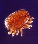
- More commonly known as the honeybee mite
- Notifiable disease in the UK
- Oval
- 1-1.5mm in length
- Eggs are laid in the hive and develop with the brood cells
Pathogenesis
- Blood-sucking
- Weakens adult bees
- Damages growing larval bees resulting in deformities
Control
- Acaricidal strips hung between combs
- Destroy colonies in apiary (if isolated outbreak)
- Monitor mite numbers and treat if widespread


