Avian Respiration - Anatomy & Physiology
Introduction
The avian respiratory system contains some fundamental differences to the mammalian system.
Avian Nasal Cavity and Oropharynx
The nostrils of the bird, which lead into the nasal cavity, may have a flap of horn to protect them, known as the Operculum. The Oral Cavity and the Nasal Cavity of the bird are interconnecting via a slit in the hard palate called the choana. Birds lack a soft palate.
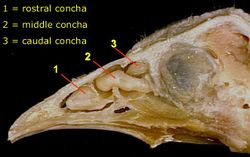
There are rostral, middle and caudal conchae arising from the lateral wall, filling part of the nasal cavity. The rostral conchae in the vestibular region are lined with stratified squamous epithelium. The middle conchae in the respiratory region are lined with respiratory epithelium. The caudal conchae in the olfactory region are lined with olfactory epithelium. The infraorbital sinus is a triangular cavity under the skin, rostroventral to the eye. Some marine birds have a salt gland (nasal gland) which excretes sodium.
Avian Larynx
The larynx is on the floor of the oropharynx supported by cricoid and paired arytenoid cartilages which are different in structure to those in mammals. There is no epiglottis and there are no vocal folds - birds vocalise using a syrinx.
Avian Trachea
The avian trachea is composed of tightly stacked cartilages which are shaped similarly to signet rings. They are complete with no dorsal space as in the mammalian trachea and overlap considerably. The trachea can be palpated on the right side of the neck; it runs alongside the oesophagus. The trachea is lined with respiratory epithelium. The trachea bifurcates into two main bronchi as in mammals. The syrinx is formed by this terminal part of the trachea.
Avian Lungs
Avian lungs are relatively compact, with a bird's lungs being approximately 50% as large as the lung of a mammal of a similar size. The lungs are unlobed and do not have the capacity to expand due to the close arrangement between the finite gas exchange structures, i.e.the air capillaries and blood capillaries and scanty connective tissue. The lungs are positioned in the craniodorsal region of the body, and are deeply indented by both the thoracic vertebrae and ribs. Birds do not have a pleural cavity as the lungs do not expand, thus the membranes are not necessary. One primary bronchus from the trachea enters each lung, narrowing as it travels through, and communicates with the abdominal air sac. This bronchus gives off branches as it travels through the lung, known as secondary bronchi. Each of these gives off a further 400-500 parabronchi in the walls of which, gaseous exchange takes place.
Air Sacs
Birds lack a diaphragm, and their thoracic and abdominal cavities are continuous. The bird has a number of thin walled, easily distensible air sacs which can extend to approximately 10x the volume of the lungs. Theye are present within body cavities, and extend into some specific bones to take the place of the bone marrow. This has the added function of reducing the weight of the bone, as they are essentially filled with air. The air sacs create unidirectional flow of air to maximise oxygen extraction and reduce heat production during flight.
The chicken has 8 air sacs:
- Cervical - extends within the cervical and thoracic vertebrae.
- Clavicular - lies within the thoracic inlet, surrounding the heart, and within the humerus in the forelimb.
- Cranial Thoracic (x2) - these are ventral to the lungs.
- Caudal Thoracic (x2) - located between the body wall and the thoracic air sacs.
- Abdominal (x2) - these are the largest air sacs and fill the caudodorsal region of the abdomen, in contact with small and large intestines, kidneys and reproductive organs. In addition, these airsacs utilise space within the acetabulum and synsacrum.
The cervical, clavicular and cranial thoracic air sacs form one functional group - cranial and the caudal thoracic and abdominal air sacs forming another, caudal functional group. The air sacs have a vital role in ventilation, but do not have the capacity for gaseous exchange.
Avian Ventilation
Ventilation in birds is strikingly different to that of mammals in that air flows through the lungs in the same direction during both inspiration and expiration. In addition, both the intaking of air and the expelling of air are active processes, requiring muscle contraction.
During inspiration, the ribs are drawn forwards and the sternum lowered, the caudal air sacs receiving fresh air. Simultaneously, the Cranial air sacs receive air which was inhaled at the previous inhalation which is drawn from the lungs, this air has lost much of its oxygen content.
During expiration, the sternum is drawn caudal and dorsal, the air sacs are compressed, air from the caudal air sac passes through the lungs, while the air in the cranial air sac leaves via the trachea. Thus, oxygenated air passes through the lungs on both inspiration and expiration.
Avian Gas Exchange
Avian Gas exchange takes place not in alveoli, as in mammals, but within air capillaries which are extensions of the parabronchial lumen. They are an interconnecting network of loops, and closely intertwine with blood capillaries. The air capillaries and blood capillaries are arranged so that flow is crosscurrent. This makes the gaseous exchange, which occurs from one to the other, extremely efficient.
References
Dyce, K.M., Sack, W.O. and Wensing, C.J.G. (2002) Textbook of Veterinary Anatomy. 3rd ed. Philadelphia: Saunders.
Sjaastad, O.V., Hove, K. and Sand, O. (2004) Physiology of Domestic Animals. Oslo: Scandinavian Veterinary Press.
Error in widget FBRecommend: unable to write file /var/www/wikivet.net/extensions/Widgets/compiled_templates/wrt69393be5cb4ff5_87687270 Error in widget google+: unable to write file /var/www/wikivet.net/extensions/Widgets/compiled_templates/wrt69393be5d50e21_48973303 Error in widget TwitterTweet: unable to write file /var/www/wikivet.net/extensions/Widgets/compiled_templates/wrt69393be5dcb7d3_16280018
|
| WikiVet® Introduction - Help WikiVet - Report a Problem |
