Penis - Anatomy & Physiology
Introduction
The penis is the male copulatory organ. It is formed from three parts; two Corpora cavernosa, comprising of cavernous tissue and a connective tissue sheath the tunica albuginea, and the single Corpus Spongiosum which contains the urethra encased in a vascular tissue sleeve. There are two types of penis: the musculovascular and fibroelastic penis.
Structure
Corpus Cavernosum
The corpus cavernosum is made up from the paired columns of cavernous tissue surrounded by connective tissue known as the crura of the penis or corpora cavernosa.
- In the dog the distal corpus cavernosum is transformed into bone to form the Os penis which plays an important part in the dog achieving intromission with the bitch during copulation.
Corpus spongiosum
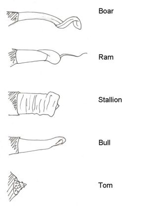
The corpus spongiosum is a vascular tissue sleeve surrounding the urethra. It commences at the bulb of the penis as an enlargement of the spongy tissue of the pelvic urethra. At the end of the penis the corpus spongiosum expands over the distal end of the corpus cavernosum to form the Glans penis, bringing the urethra to the extremity of the penis. There is a great variation in the morphology of the glans penis between species which often correspond to the female tract morphology. For example the boars “corkscrew” glans penis corresponds to the many interdigitating prominences of the sow’s cervix. The glans penis is highly populated with sensory nerves.
Prepuce
The prepuce is the skin sheath that conceals the penis when it is flaccid and is formed by an invagination of the abdominal skin. The prepuce is hairless and contains many smegma secreting glands important for lubrication between the shaft of the penis and the prepuce during copulation. Within the prepuce are varying amounts of striated muscle fibres; the cranial prepucial muscles responsible for retracting the prepuce and the caudal prepucial muscles responsible for protracting the prepuce.
Glans Penis morphology
- Bull – slightly spiralled end
- Boar – corkscrew shaped with a left hand thread
- Ram – large extension of the urethral process
- Stallion – Mushroom shaped with slight protrusion of the urethral process
- Dog – substantial glans penis which is divided into the bulbus glandis found proximally and the pars longa glandis found distally
- Tom cat – cone shaped with Keratinised Papillae, directed caudally
Types of Penis
Fibroelastic
Found in:
- Bull, Boar, Ram, Deer
- Contain large amounts of connective tissue and elastic fibres but limited erectile tissue.
- Contain a sigmoid flexure
- They are encased by a non-expandable connective tissue sheath called the tunica albuginea. Therefore, erection only results in increased length of penis and no increase in diameter of the penis. However, most of the increase in penile length is actually due to the straightening of the sigmoid flexure.
- The cavernous tissue contains small blood spaces which means that only a small increase in blood to the penis is require to achieve erection.
Musculovascular
Found in:
- Man, Stallion, Dog, Tom Cat
- This penis structure contains a lot of erectile tissue and little connective tissue so during erection there is both an increase in length and diameter of the penis.
- The cavernous tissue contains large blood spaces divided by thin septa. Therefore, a relatively larger volume of blood is required to achieve erection.
Muscles associated with the penis
Bulbospongiosus
- A single muscle that covers the root and ventral surface of the penis as well as the bulbourethral glands (link to glands page)
- The function of this muscle is to empty the extrapelvic urethra of sperm in a similar way to the urethralis muscle emptying the pelvic urethra.
Ischiocavernosus Muscles
- Paired muscles located at the root of the penis.
- Connect the penis to the ischial arch of the pelvis.
Retractor penis Muscles
- Paired muscles originating on the caudal vertebrae and inserting on the ventrolateral surfaces of the penis.
- Maintains the sigmoid flexure of the fibroelastic penis when the muscles are contracted.
- When the muscles are relaxed the penis protrudes through the prepuce as the sigmoid flexure unbends.
Vasculature
The artery of the penis is a direct branch off the internal pudendal artery. It splits into three branches:
- Artery of the bulb – supplies the corpus spongiosus
- Deep artery of the penis – supplies the corpus cavernosum
- Dorsal artery of the penis – supplies the glans penis
The prepuce covering the flaccid penis is supplied by anastamosis between the external pudendal artery and the artery of the penis.
Innervation
- Mostly parasympathetic from the paired pudendal nerves
- The glans penis and internal lamina of the prepuce are heavily infiltrated by sensory nerve endings responsible for stimulating ejaculation (link to ejaculation)
Lymphatics
Lymph from the penis and prepuce drains into the superficial inguinal lymph nodes.
Webinars
Feline Lower Urinary Tract Disease (FLUTD) - focussing on causes and management
| Penis - Anatomy & Physiology Learning Resources | |
|---|---|
 Test your knowledge using drag and drop boxes |
Test your knowledge of penis structure in different species |
 Multiple choice quizzes |
Male Reproductive Anatomy Quiz |
 Selection of relevant videos |
Equine penis potcast Reproductive tract of the boar Reproductive tract of the dog potcast |
Error in widget FBRecommend: unable to write file /var/www/wikivet.net/extensions/Widgets/compiled_templates/wrt675a790fb589e0_25614541 Error in widget google+: unable to write file /var/www/wikivet.net/extensions/Widgets/compiled_templates/wrt675a790fbc4c61_35104943 Error in widget TwitterTweet: unable to write file /var/www/wikivet.net/extensions/Widgets/compiled_templates/wrt675a790fc29230_88264880
|
| WikiVet® Introduction - Help WikiVet - Report a Problem |
