Cell Growth Disorders
Jump to navigation
Jump to search
| This article has been peer reviewed but is awaiting expert review. If you would like to help with this, please see more information about expert reviewing. |
|
|
Atrophy
- Atrophy is a decrease in the size of the cells and organ, occurring after the organ has reached normal size.
Appearance
- Atrophic tissues and organs appear smaller and perhaps paler than usual.
- Microscopy of atrophic tissue shows:
- Cells of a smaller size.
- Inactive appearance.
- A relative increase in the supportive connective tissue.
Causes
- Starvation
- Malabsorption
- Compression
- E.g by a nearby lesion.
- Immobilisation
- Immobilisation of a limb results in atrophy of the muscles.
- Denervation
- Lack of trophic hormones
- Chronic inflammation
- May be idiopathic.
Examples
Serous Atrophy of Adipose Tissue
- Also known as gelatinous atrophy of adipose tissue.
- The fat becomes transparent, watery and severely depleted.
- Occurs as a result of severe debilitation and weight loss.
Brown Atrophy
- A senile change in muscles where they appear brownish rather than reddish in colour.
- Due to the intracytoplasmic accumulation of lipofuscin within the muscle fibres.
- The "wear and tear" pigment.
Hypertrophy
- Hypertrophy is an increase in the size of an organ due to an increase in size of the individual cells.
- The organ also gains in weight.
Types
Functional Hypertrophy
- Occurs in response to:
- An increased physiological need.
- E. g. muscles of the heart and limbs in training.
- An increased demand because of organ dysfunction.
- E.g in cardiac hypertrophy due to a progressively failing heart.
- An increased physiological need.
Compensatory Hypertrophy
- Occurs when one of a paired organ is damaged or lost.
- E.g. the kidney.
Obstructional Hypertrophy
- Hollow organs may become thickened around an obstruction.
- E. g. intestine, bladder, gall bladder.
Hormonal Mediated Hypertrophy
- Anabolic steroids produce hypertrophy of muscle.
- Thyroid hormones have a general hypertrophic effect on tissues.
- Increase protein synthesis within them.
- The heart can become quite hypertrophied in thyroid excess.
- Commonly seen in older cats which often develop hyperthyroidism.
Hypoplasia
- Hypoplasia is a reduction in the size of cells and tissues,.
- Due to a failure to grow to normal size.
- Ranges from mild hypoplasia to almost complete absence.
- Almost complete absence is also called vestigial or rudimentary.
- Aplasia and agensis refer to complete absence of tissue.
- Generally refer to the gross appearance rather than the microscopic appearance.
- Some rudimentary tissue can be seen if searched for carefully.
- Generally refer to the gross appearance rather than the microscopic appearance.
Hyperplasia
- Hyperplasia is an increase in the size of an organ due to an increase in the numbers of cells present within it.
- Hypertrophy and hyperplasia may occur concurrently.
- The hyperplastic response stops when the inciting agent ceases.
- Hyperplastic tissue is more prone to injury by chemicals, and also may be more prone to undergo neoplastic change in some cases.
Causes
- Hormonal stimulation.
- Parathyroid hyperplasia in chronic renal failure.
- Prostatic hyperplasia in older dogs.
- Hyperplasia may also occur as a regenerative response to
- Irritation
- Cell loss
- Injury
Gross Appearance
- Hyperplastic nodules can be seen in a variety of organs in older dogs and cats.
- Particularly the thyroid in cats, and the spleen and liver of dogs.
Histological Appearance
- Hyperplasia is characterised by an increase in the numbers of cells.
- Mitotic activity is not always seen.
- There is some increase in cellular basophilia.
- The cells are well differentiated and tissue structure is normal.
Metaplasia
- Metaplasia is a transformation of one type of tissue into another.
- Occurs solely in:
- Connective tissue
- The metaplastic change id to cartilage and bone in damaged tissue.
- Caesarean scars in the pig are especially prone to osseous metaplasia.
- Epithelium
- Squamous metaplasia of cuboidal or columnar epithelium is quite common.
- Seen in the prostate of dogs under the influence of oestrogens.
- Oestrogens are present in Sertoli cell tumours and in Vit. A deficiency.
- The most striking example is the squamous metaplasia of the oesophageal glands in the chicken.
- Seen in the prostate of dogs under the influence of oestrogens.
- Squamous metaplasia of cuboidal or columnar epithelium is quite common.
- Connective tissue
- Mixed tumours of the mammary gland of the dog are so called because there is proliferation of both the glandular element and the surrounding myoepithelium.
- The myoepithelium can transform into cartilage and bone.
- Some regard this as a metaplastic change.
- The bone formed may even show marrow formation within the spaces between the bony trabeculae.
- The myoepithelium can transform into cartilage and bone.
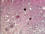
Dysplasia
- Dysplasia is abnormal growth within a tissue.
- The normal arrangement and pattern of the tissue may be lost.
- The most common example is the renal dysplasia in some breeds of dogs.
- Causes fibrous tracts.
- Because of the reduction in functional tissue, gives a predisposition to renal failure early in life in the more severely affected cases.
Anaplasia
- Anaplasia is a marked and irreversible loss of cellular differentiation with return to a more primitive state.
- Afeature of highly malignant tumours.
- There is no pattern, just a diffuse sheet of cells.
- Both cells and nuclei are of differing sizes.
- There are prominent nucleoli in some nuclei.
