Panosteitis
Introduction
Panosteitis is a spontaneous, self-limiting inflammatory disease of young, rapidly growing large or giant dogs. 75% of cases are seen in German Shepherd Dogs between 5 and 12 months of age. It is much more common in males than in females.
It most commonly causes forelimb lameness, and affects the diaphyses of the proximal ulna, central radius, proximal and distal humerus, central femur and proximal tibia. Less commonly, the metacarpal bones and pelvis may be affected.
The exact aetiology is unknown, but genetics, stress, infection, and metabolic or autoimmune causes have been suspected.
The pathophysiology of the disease is characterised by intramedullary fat necrosis, excessive osteoid production, and vascular congestion. Endosteal and periosteal bone reactions occur.
Clinical Signs
Dogs usually present with a cyclic or shifting long bone lameness. The lameness is often episodic and can affect different bones at different times.
There is marked pain when firm digital pressure is applied to the affected bones.
Affected dogs can be inappetent and pyrexic.
Diagnosis
Panosteitis can be suspected if long bone pain occurs in dogs with the right signalment.
Radiography does not always diagnose the condition at first presentation as radiographic signs lag behind clinical signs and are often subtle.
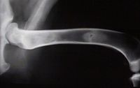
The characteristic radiological appearance of panosteitis is an increase in bone density in the middle of a long bone, almost like a thumb print.
The radiographic signs can be divided into three phases:
- The early phase is characterised by a granular increase in intramedullary radiodensity with loss of corticomedullary distinction.
- The middle phase is characterised by a change in the intramedullary density to a patchy or mottled appearance. The density of the medulla may appear similar to that of cortical bone at this stage. These changes may be centered around the nutrient foramen. Periosteal and endosteal new bone formation resulting in cortical thickening develops during this phase.
- In the late phase, the appearance of the medullary canal returns to normal. The thickened cortices and accentuated trabecular pattern are the last to resolve.
Such lesions may be present in a single bone or in several bones. Repeat radiographs are helpful to assess the progression of the condition through the phases. Sometimes a definitive radiological diagnosis is never achieved because the clinical signs disappear before further radiographs are taken.
Differential diagnoses would include: osteomyelitis and hypertrophic osteodystrophy.
Treatment
Panosteitis is treated conservatively using [[NSAIDs[[ during episodes of lameness and pain, with strict exercise restriction.
Recurrent episodes can be expected and several courses of analgesia may be necessary. The disease is usually self-limiting in one to several months.
Owners may be taught to recognise signs of early pain and can administer short courses of NSAIDs on an 'as needed' basis.
The prognosis is excellent and the disease is rarely seen past the age of 20 months.
Excessive dietary supplementation in young, large-breed growing dogs should be avoided.
| Panosteitis Learning Resources | |
|---|---|
 Test your knowledge using flashcard type questions |
Small Animal Emergency and Critical Care Medicine Q&A 18 |
References
Hill, P. (2011) 100 Top Consultations in Small Animal General Practice John Wiley and Sons
Lewis, D. (1998) Self-assessment colour review of small animal orthopaedics Manson Publishing
Merck and Co (2008) The Merck Veterinary Manual Merial
| This article has been peer reviewed but is awaiting expert review. If you would like to help with this, please see more information about expert reviewing. |
