Difference between revisions of "Amyloidosis"
Michuang0720 (talk | contribs) |
|||
| (9 intermediate revisions by 4 users not shown) | |||
| Line 1: | Line 1: | ||
| − | == | + | ==Introduction== |
| − | Amyloid infiltration occurs in all species - | + | Amyloid infiltration occurs in all species - amyloid is an inert substance that becomes deposited under the endothelium and basement membranes of a variety of tissues, notably the renal glomeruli, |
[[Pancreas - Anatomy & Physiology#Endocrine|Islets of Langerhans]] in the [[Pancreas - Anatomy & Physiology|pancreas]] and the [[Liver - Anatomy & Physiology|liver]] (between the sinusoidal reticulum and the hepatic cords). | [[Pancreas - Anatomy & Physiology#Endocrine|Islets of Langerhans]] in the [[Pancreas - Anatomy & Physiology|pancreas]] and the [[Liver - Anatomy & Physiology|liver]] (between the sinusoidal reticulum and the hepatic cords). | ||
==Causes== | ==Causes== | ||
| − | May be a primary condition or appear secondary to some chronic tissue destructive process | + | May be a primary condition or appear secondary to some chronic tissue destructive process such as: |
*an infectious process elsewhere in the body | *an infectious process elsewhere in the body | ||
| − | * | + | *sustained antigenic stimulation, eg repeated injections with an antigenic substance or production of excessive antibody by leukocytes |
==Gross Pathology== | ==Gross Pathology== | ||
| + | Affected organs will apear: | ||
*pale (greyish waxy appearance) | *pale (greyish waxy appearance) | ||
| − | *enlarged | + | *enlarged with rounded edges |
| − | |||
*firm | *firm | ||
*very prone to rupture | *very prone to rupture | ||
| Line 19: | Line 19: | ||
*shows 'apple-green' fluorescence under polarised light after staining with Congo Red | *shows 'apple-green' fluorescence under polarised light after staining with Congo Red | ||
| − | ==Arterial== | + | ==Arterial Dissemination== |
| − | Amyloid is an eosinophilic, homogenous, hyaline material. Due to its beta-pleated-sheet structure it is almost insoluble. Amyloid may be: | + | Amyloid is an eosinophilic, homogenous, hyaline material. Due to its beta-pleated-sheet structure it is almost insoluble. Amyloid may be present in one of two forms: |
*'''AA''': Serum amyloid A, alpha-2 globulin. | *'''AA''': Serum amyloid A, alpha-2 globulin. | ||
*'''AL''': Derived from immunoglobulin light chains. | *'''AL''': Derived from immunoglobulin light chains. | ||
| Line 26: | Line 26: | ||
Disease may be truly idiopathic (dogs and cats) or may be secondary to another disease process, often chronic inflammation or neoplasia. Chronic antigenic stimulation induces the overproduction of AA protein which may become deposited throughout the body. | Disease may be truly idiopathic (dogs and cats) or may be secondary to another disease process, often chronic inflammation or neoplasia. Chronic antigenic stimulation induces the overproduction of AA protein which may become deposited throughout the body. | ||
| − | Deposits | + | Deposits can be found in: |
*Renal vessels and glomeruli. | *Renal vessels and glomeruli. | ||
*Splenic white pulp. | *Splenic white pulp. | ||
| Line 35: | Line 35: | ||
Affected organs are non-functional and appear waxy and pale. | Affected organs are non-functional and appear waxy and pale. | ||
| − | |||
| − | [[ | + | *Amyloid is an insoluble protein that can accumulate in the kidney and compress the glomerulus, interfering with its normal function. |
| + | *Hypoproteinaemia and nephrotic syndrome can result due to substantial protein loss in the urine. | ||
| + | *Causes can be idiopathic or associated with underlying chronic inflammatory conditions. | ||
| + | '''Gross pathology''' | ||
| + | *Kidneys are firm, enlarged, and pale. Affected glomeruli may be seen as yellowish spots in the cortex. | ||
| + | *Amyloid can be visualised by treating the freshly cut surface of the kidneys with iodine. | ||
| + | |||
| + | '''Histopathology''' | ||
| + | *Amyloid stains pink with congo red. | ||
| + | *Presence of a pink, homogenous material replacing the epithelium and endothelium of the glomerulus. | ||
| + | <center><gallery> | ||
| + | Image:Congo red.jpg|'''Amyloidosis Stained with Congo Red''' <br> Susan Rhind, University of Edinburgh | ||
| + | Image:Amyloidosis_histology.jpg|'''Amyloidosis Histology''' <br> Susan Rhind University of Edinburgh | ||
| + | |||
| + | </gallery></center> | ||
| + | |||
| + | |||
| + | |||
| + | |||
| + | {{Learning | ||
| + | |Vetstream = [https://www.vetstream.com/canis/search?s=amyloidosis Amyloidosis] | ||
| + | |flashcards=[[Vascular Pathology Flashcards|Vascular Pathology]] | ||
| + | }} | ||
| + | |||
| + | |||
| + | |||
[[Category:Liver_-_Storage_Diseases]] | [[Category:Liver_-_Storage_Diseases]] | ||
[[Category:Cardiovascular_System_-_Degenerative_Pathology]][[Category:Cardiovascular_System_-_Vascular_Pathology]][[Category:Arterial_Pathology]] | [[Category:Cardiovascular_System_-_Degenerative_Pathology]][[Category:Cardiovascular_System_-_Vascular_Pathology]][[Category:Arterial_Pathology]] | ||
| + | [[Category:Glomerular Disease]] | ||
| + | [[Category:To Do - Urinary]][[Category:Cardiology Section]] | ||
Latest revision as of 20:33, 25 June 2016
Introduction
Amyloid infiltration occurs in all species - amyloid is an inert substance that becomes deposited under the endothelium and basement membranes of a variety of tissues, notably the renal glomeruli, Islets of Langerhans in the pancreas and the liver (between the sinusoidal reticulum and the hepatic cords).
Causes
May be a primary condition or appear secondary to some chronic tissue destructive process such as:
- an infectious process elsewhere in the body
- sustained antigenic stimulation, eg repeated injections with an antigenic substance or production of excessive antibody by leukocytes
Gross Pathology
Affected organs will apear:
- pale (greyish waxy appearance)
- enlarged with rounded edges
- firm
- very prone to rupture
Microscopically
- deposition of amyloid in the space of Disse (or perisinusoidal space) which is in the liver between the hepatocytes and a sinusoid.
- shows 'apple-green' fluorescence under polarised light after staining with Congo Red
Arterial Dissemination
Amyloid is an eosinophilic, homogenous, hyaline material. Due to its beta-pleated-sheet structure it is almost insoluble. Amyloid may be present in one of two forms:
- AA: Serum amyloid A, alpha-2 globulin.
- AL: Derived from immunoglobulin light chains.
Disease may be truly idiopathic (dogs and cats) or may be secondary to another disease process, often chronic inflammation or neoplasia. Chronic antigenic stimulation induces the overproduction of AA protein which may become deposited throughout the body.
Deposits can be found in:
- Renal vessels and glomeruli.
- Splenic white pulp.
- Space of Disse.
- Coronary arteries.
- Meningeal arteries.
Affected organs are non-functional and appear waxy and pale.
- Amyloid is an insoluble protein that can accumulate in the kidney and compress the glomerulus, interfering with its normal function.
- Hypoproteinaemia and nephrotic syndrome can result due to substantial protein loss in the urine.
- Causes can be idiopathic or associated with underlying chronic inflammatory conditions.
Gross pathology
- Kidneys are firm, enlarged, and pale. Affected glomeruli may be seen as yellowish spots in the cortex.
- Amyloid can be visualised by treating the freshly cut surface of the kidneys with iodine.
Histopathology
- Amyloid stains pink with congo red.
- Presence of a pink, homogenous material replacing the epithelium and endothelium of the glomerulus.
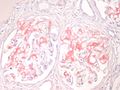
Amyloidosis Stained with Congo Red
Susan Rhind, University of Edinburgh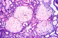
Amyloidosis Histology
Susan Rhind University of Edinburgh


| Amyloidosis Learning Resources | |
|---|---|
To reach the Vetstream content, please select |
Canis, Felis, Lapis or Equis |
Test your knowledge using flashcard type questions |
Vascular Pathology |